Origami cranes — with their elegant paper wings and distinct angular beaks — rely on symmetry in their folding pattern to achieve their graceful form. This symmetry mirrors the bilateral balance seen in real birds and enables complexity to emerge in fewer steps. Yet, for the crane’s head to stand apart from its tail, the design must break free from strict symmetry. This subtle shift allows for unique features that perfect symmetry cannot achieve.
Taking inspiration from this concept, two new studies published back-to-back in Nature (Lee et al.; Dowling et al.) from the Baker and King Labs introduce asymmetry into the design of protein nanocages — tiny structures with enormous potential for vaccines and drug delivery. These nanocages are like advanced origami creations, built from many intricately folded protein parts.
Read the authors’ summary of this work, including how the two papers relate, here.
Four-component protein nanocages designed by programmed symmetry breaking
Authors:Sangmin Lee, Ryan D. Kibler, Green Ahn, Yang Hsia, Andrew J. Borst, Annika Philomin, Madison A. Kennedy, Buwei Huang, Barry Stoddard, David Baker
Hierarchical design of pseudosymmetric protein nanocages
Authors:Quinton M. Dowling, Young-Jun Park, Chelsea N. Fries, Neil C. Gerstenmaier, Sebastian Ols, Erin C. Yang, Adam J. Wargacki, Annie Dosey, Yang Hsia, Rashmi Ravichandran, Carl D. Walkey, Anika L. Burrell, David Veesler, David Baker & Neil P. King
Sangmin Lee, PhD
Ryan Kibler, PhD
Quinton Dowling, PhD
Building larger, more complex structures
“For the last decade, we were limited to building protein nanocages with strict symmetry,” said Neil King, associate professor of biochemistry at UW Medicine. “But in these two papers, we’ve used a principle called pseudosymmetry to design cage-like protein assemblies that are considerably larger than anything we’ve made before. These new types of nanocages could someday serve as highly advanced containers, packaging medicines and delivering them to precise locations in the body, which could make treatments safer and more effective.”
Cryo-EM structure of an octohedral protein nanocage with 96 subunits built from four unique proteins.
Cryo-EM structure of an icosahedral protein nanocage with 240 subunits built from four unique proteins.
The King Lab has pioneered the design and use of custom protein nanoparticles as vaccine components, leading them to develop the world’s first computationally designed protein medicine with partners at UW Medicine.
Beyond strict symmetry
The scientists drew inspiration from viruses, which are masters of building complex structures with subtle breaks in symmetry. Using pseudosymmetry — where similar but not identical parts combine to form larger assemblies — the team computationally designed and experimentally verified an array of custom nanocages. These included structures with 240, 540, and even 960 protein subunits — by far the largest protein nanocages ever created to date. The largest structure, measuring nearly 100 nanometers across, has a volume 90 times greater than the well-studied AAV viral capsid.
These new nanocages are not just bigger but also vastly more complex. One assembly contains four distinct protein chains and six unique protein-protein interfaces, all designed entirely from scratch.
“This work revealed secrets of the formation of large virus capsid structures and paved ways to intentionally design similar structures for biomedical applications such as cell targeting and gene delivery,” said Sangmin Lee, a co-lead author and former Baker Lab postdoctoral scholar who is now an assistant professor at Pohang University of Science and Technology (POSTECH).
To achieve this leap, the teams also embraced quasisymmetry — the use of identical protein parts that adopt different shapes depending on their local environment. Designing these shifts in form required extraordinary computational precision, resulting in nanocages with entirely novel architectures.
“Every time I saw one of these new assemblies on the electron microscope display, I had to pause and admire it,” said Ryan Kibler, co-lead author and postdoctoral researcher in the Baker Lab. “Their shapes are so distinct and unusual that it’s obvious they are man-made. This is especially true for the tetrahedral and octahedral types, which, to our knowledge, have never been observed in nature.”
These supersized nanocages represent a major step forward in protein design. Pushing beyond the limits of symmetry opens the door to creating more sophisticated molecular tools that more closely resemble the elegant functions seen in living systems. With their potential as vaccine scaffolds and containers for targeted drug delivery, these intricate assemblies may one day reshape how we treat disease and build nanoscale technologies.
Led by the Institute for Protein Design, this research included collaborators from the the IPD Core R&D Labs, the Stoddard Lab at the Fred Hutchinson Cancer Research Center, the Wysocki Lab at the Ohio State University, and the Veesler Lab at UW Medicine.
Researchers at the Institute for Protein Design have developed a new AI-driven molecular design tool that generates completely novel bioactive peptides. The ability to create such molecules by computation alone may accelerate drug development.
RFpeptides leverages deep learning to design ring-shaped peptides called macrocycles that bind to disease-associated proteins using only the structure or sequence of a target. This is a departure from traditional peptide discovery methods, which often require extensive screening of vast peptide libraries to identify potential binders.
Introduced as a preprint, RFpeptides was developed in the laboratories of three IPD faculty members: Gaurav Bhardwaj, Frank DiMaio, and 2024 Nobel laureate David Baker. Trainees Stephen Rettie, David Juergens, and Victor Adebomi led this project.
Victor Adebomi, PhD
David Juergens, PhD
Stephen Rettie
“RFpeptides extends the AI revolution in biology to the important challenge of peptide design. We hope it will help researchers create peptide-based medicines for a variety of diseases that have no good treatment options today,” said Gaurav Bhardwaj, an assistant professor of medicinal chemistry at the UW School of Pharmacy.
The University of Washington has licensed RFpeptides to Vilya, an IPD spinout company. Bhardwaj and Baker are co-founders, advisors, and shareholders of the company.
What is RFpeptides?
RFpeptides in action: Artist’s depiction of a colorful macrocycle being generated by RFpeptides.
RFpeptides is a software tool for designing bioactive peptides with precise 3D structures. Peptides are molecules composed of only a few amino acid building blocks. When the first and last of these building blocks link together, the peptide forms a cyclic structure. This often makes the peptide more resistant to degradation and can confer other biochemical benefits, such as a more rigid structure for higher affinity target binding. The precise nature of these binding interactions can activate or deactivate a target protein and thus drive a biological effect.
How does it work?
RFpeptides builds upon the success of RFdiffusion and introduces key innovations tailored for the specific challenges of macrocyclic peptide design. Pioneered at the IPD, both tools draw inspiration from popular AI image generators that use diffusion models to synthesize new images based on user prompts. With RFdiffusion, a diffusion model sculpts clouds of disconnected amino acids into plausible biochemical structures. To create RFpeptides, the team modified the open-source tool to ensure the first and last amino acids in a designed molecule could form a chemical bond.
“By expanding the structure modeling capabilities of RoseTTAFold2 and harnessing ProteinMPNN for sequence design, we’ve created a peptide design pipeline that’s both computationally efficient and incredibly accurate,” said associate professor of biochemistry Frank DiMaio.
Testing RFpeptides
To demonstrate that RFpeptides can produce functional binding peptides for a range of challenging targets, the team selected four proteins implicated in hospital-derived bacterial infection, cancer, and other cellular processes important to human health. They synthesized and tested over a dozen designed macrocycle binders, identifying high-affinity interactions with each target.
“The highlight for me was that we successfully produced a high-affinity binder for a target with no known structure. Starting from just the target’s amino acid sequence, we predicted its structure using AlphaFold 2 and RoseTTAFold 2, designed peptides to bind those predicted structures, and ended up solving the first structure of the pathogenic protein Rhombotarget A,” said co-lead author and graduate student Stephen Rettie.
Adapted from Figure 4: Accurate de novo design of a high-affinity cyclic peptide binder against the predicted structure of RbtA from A. baumannii. (A) The macrocycle shown in purple was designed to bind to the bacterial protein target shown in gray. (B) Binding experiments revealed that the molecules interact with high affinity. (C) A high-resolution X-ray structure of the complex reveals that the binding interaction was designed with atomic accuracy.
“This is quite a neat display of the robustness and generalization capacity these generative models gain during pretraining” said co-lead author David Juergens. “The implications for drug design are really exciting as macrocycles can be customized in many ways that normal peptides and proteins cannot.”
The development of RFpeptides marks another step in leveraging AI to solve complex challenges in biology. While initially tested as a drug design tool, RFpeptides could also be used to create diagnostic reagents and other custom peptides for research challenges beyond medicine.
Computational protein design, once considered impossible, has blossomed into a transformative research field with implications for medicine, sustainability, and beyond. In a recent webinar hosted by David Baker, three pioneers in the field—Bill DeGrado, Steve Mayo, and Brian Kuhlman—shared their insights on the origins, evolution, and future of protein design.
David Baker has been awarded the 2024 Nobel Prize in Chemistry for computational protein design. Baker is a professor of biochemistry, Howard Hughes Medical Institute investigator, and director of the Institute for Protein Design at the University of Washington School of Medicine. The prize is shared with Demis Hassabis and John Jumper of Google DeepMind for their contributions to protein structure prediction.
Proteins are life’s most important molecules. Found in every organism, they perform nearly all biological processes — from cell communication and growth to immunity and replication. And in the words of the Nobel committee, Baker “has succeeded with the almost impossible feat of building entirely new kinds of proteins.”
Baker’s research combines computational and laboratory techniques to create custom proteins with unprecedented precision. The Baker Lab pioneered the field by developing much of the world’s most popular and effective software for computational protein design, including the Rosetta program. The lab also succeeded in creating the first protein with a novel 3D shape, an inert molecule called Top7. It has since produced several thousand new proteins, including molecules that neutralize viruses, target cancer cells, and even serve as catalysts for chemical reactions. Baker’s research also contributed to the development of the world’s first computationally designed protein medicine, a protein-based vaccine for COVID-19 pioneered by colleagues at UW Medicine.
“We have entered an era where we can not only understand biological systems but also create new ones,” Baker explained. “By designing proteins not found in nature, I believe we will solve many urgent challenges across medicine, technology, and sustainability. This prize is a testament to the collective efforts of the many hundreds of brilliant scientists whom I have had the pleasure to work with, and it is an honor to be part of this exciting moment in science.”
Recently, artificial intelligence has been tapped by Baker and others to predict and design protein structures with unprecedented accuracy and speed. This has greatly expanded scientists’ ability to model and design the building blocks of life. As the 2024 Nobel Prize in Chemistry makes clear, no other domain of science has been more transformed by AI than protein research.
To date, Baker has published more than 640 peer-reviewed research papers, been awarded over 100 patents, and co-founded 21 biotechnology companies. Ninety of his trainees have gone on to independent faculty positions. His dedication to open science has fostered a collaborative community of researchers worldwide, and he has ensured that the most advanced tools and insights developed through his work are shared freely to accelerate scientific discovery.
Joining the ranks of Nobel Laureates is a testament to Baker’s dedication, innovation, and the far-reaching implications of his work and that of his co-awardees with whom this honor is shared. We extend our heartfelt congratulations to these remarkable scientists and remain committed to nurturing world-changing research.
Today marks a historic milestone for modern science. As researchers, innovators, and communities worldwide celebrate this remarkable achievement, we look forward to the groundbreaking discoveries and life-changing solutions that still lie ahead — solutions made possible by the visionary work that has earned David Baker a Nobel Prize.
This summer, the Institute for Protein Design hosted 18 undergraduate students from across the world as visiting researchers. For 10 weeks, they experienced life in a modern research environment, learned how to conduct their own experiments, and collaborated with mentors and peers to advance significant research projects.
Training across disciplines
Protein design is an interdisciplinary field that blends ideas from computer science, biology, chemistry, and physics, meaning there’s no typical background for researchers here. This year, our cohort included students majoring in ten different disciplines, each bringing a unique perspective to their summer research.
“I came to the IPD having only explored a very specific field of research completely unrelated to protein design, and I had no background in coding, so I was quite far out of my comfort zone. I always figured that I would have no interest in computation, but this experience completely changed that for me; I quickly realized how powerful computation is in biology.”
Hannah Stewart
University of Michigan (biophysics)
2024 IPD Summer Undergraduate Researchers
IPD Happy Hour with David Baker
POV when summer’s over
Advanced research
With projects rooted in each IPD Member Lab, the students tackled a wide range of research topics, from vaccine design to software development. They arrived with varying levels of research experience and left with a deepened passion for discovery, eager to apply their newfound insights in their home labs or in future research opportunities.
“Working on designing protein binders targeting cancerous peptides was an incredible experience, and the coolest part was seeing the potential impact of my work in real time. Despite having no prior wet lab experience, my mentors, Nathan Greenwood and Julia Bonzanini, were incredibly supportive and helped me get more comfortable with the techniques. At the same time, diving into advanced computational tools was both challenging and rewarding, pushing me to explore areas outside of my comfort zone. I’m looking forward to applying my experience here to further research in precision medicine.”
Jazmin Sharp
Towson University (applied mathematics)
Forming connections
Starting on day one, students were invited to join research subgroups, participate in journal clubs, and attend seminars, becoming completely immersed in research at the IPD.
“I was amazed by the number of experts in different fields you can talk to on a daily basis, creating a perfect interplay between wet lab research and machine learning. This is a place where a coffee chat can teach you more than a dozen papers. I also loved the openness of the institute, the opportunity to meet new people every day, and the eagerness of everyone to help you succeed.”
Jie Chen
University of Texas at Dallas (computer science)
“For any future interns, working at the IPD will allow you to learn about the newest advancements to the world of protein design while interacting with scientists who truly love what they do. I am so thankful for such a welcoming environment that allowed me to learn the ins and outs of computational and experimental work within this field.”
Mariah Culpepper
Duke University (biology and computer science)
“Everyone was so friendly and open to any questions, which was incredibly valuable. Participating in happy hours, chocolate hours, and chatting in the kitchen allowed me to deepen my relationships, hear about their research, and enjoy casual conversations, creating wonderful memories. And last but not least, my mentor has been the best mentor I’ve ever had! Thanks to him, I was able to fully enjoy my research this summer.”
Arisa Uchida
University of Tokyo (education)
Sharing their work
Participants had two opportunities to present their undergraduate research projects at the end of the program. With guidance from experienced scientists, they created research posters for RosettaCon or the UW Summer STEM Undergraduate Research Poster Session to highlight their contributions and findings.
Yuvraj Balani (left) and his mentor Stephen Rettie at the UW Summer STEM Undergraduate Research Poster Session.
Mariah Culpepper won a poster award at RosettaCon!
Max Witwer at the UW Summer STEM Undergraduate Research Poster Session.
Nadya Lumy at the UW Summer STEM Undergraduate Research Poster Session.
Elliott Cole at the UW Summer STEM Undergraduate Research Poster Session.
Join us next summer
IPD Summer Research Program Funded: Yes Supported by: Institute for Protein Design Eligibility: Full-time undergraduate students (anywhere).
AI tools to design hinge proteins to α-helical peptides | Baker Lab Mentor: Kathryn Shelley & Cullen Demakis Mentee: Hannah Stewart, Elliott Cole & Rose Duong
De novo design of an azide-alkyne cyclase |Baker Lab Mentor: Declan Evans Mentee: Jie Chen
De novo design of antibodies and antibody-like molecules, including single chain variable fragments and/or nanobodies |Baker Lab Mentor: DeJenae See & Ellen Shrock Mentee: Max Witwer
De novo design of macrocycles and disulfide stapled peptides against disease-relevant targets |Bhardwaj Lab Mentor: Gizem Gokce & Stephen Rettie Mentee: Summer Solis & Yuvraj Belani
Designing peptide-major histocompatibility complex (pMHC) protein binders for targeting | Baker Lab Mentor: Nathan Greenwood & Julia Bonzanini Mentee: Jazmin Sharp
Developing water soluble RFdiffused associated protein scaffolds (WRAPS) for a variety of natural and de novo membrane proteins |Baker Lab Mentor: LJ Mihaljevic & Sagardip Majumder Mentee: Mariah Culpepper & Sungjai Shin
Generating high-quality protein-small molecule complexes using physics-based ligand docking for RoseTTAFold All-Atom training |DiMaio Lab Mentor: Davi Nakajima An Mentee: Nadya Annabelle Lumy & Grace Li
Designing fully de novo protein vaccines using a pathogen agnostic approach |King Lab Mentor: Sebastian Ols Mentee: Octavius Louis & Arisa Uchida
Design nanoparticles to display a mosaic of proteins on the surface |King Lab Mentor: Sanela Rankovic, Chelsea Fries & Susan Kleinfelter Mentee: Kristine Pham, Dominik Nowak, Favour Olushola & Ganga Dripaul
Special thanks to program organizers Kandise VanWormer and Madison Kennedy, PhD.
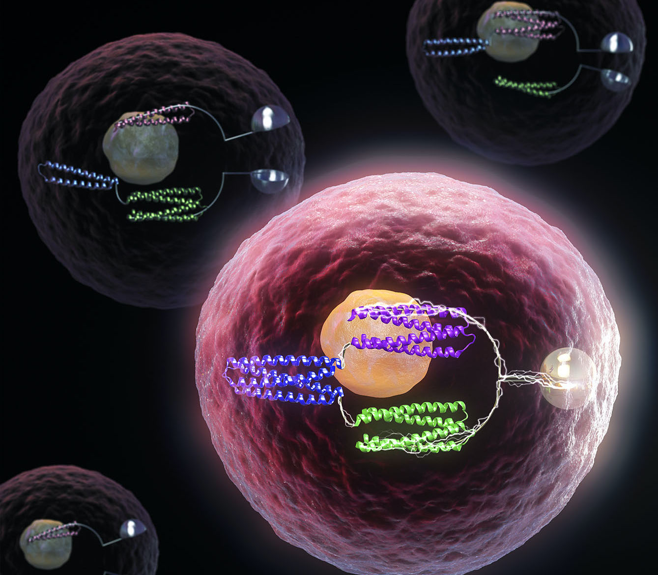
The sense of smell depends on sensitive proteins embedded in the nose. In two studies published recently in Science (1,2), a team led by the Baker Lab has shown that an effectively unlimited number of new protein sensors can now be generated with the help of AI. This is a major step toward more advanced molecular sensing technologies for use in healthcare, pollution monitoring, and more.
Biochemically speaking, smelling a rose requires two feats. First, floral compounds must bind to olfactory receptors in the nose. This molecular handshake must then produce a reliable signal, leading to the perception of smell.
In the new studies, the team used computers to create custom proteins that bind to specific compounds and transmit stable bioelectronic binding signals across lipid membranes. These sensor proteins are not derived from any found in nature. Instead, they were built from scratch and confirmed to function in the lab.
“When we started with this idea a few years ago, many people thought it was impossible,” said senior author and former Baker Lab postdoc Anastassia Vorobieva, now a group leader at the VIB-VUB Center for Structural Biology in Belgium. “Now we have shown that we can successfully design nanopore proteins with a high success rate and that they can have stable and reproducible conductance.”
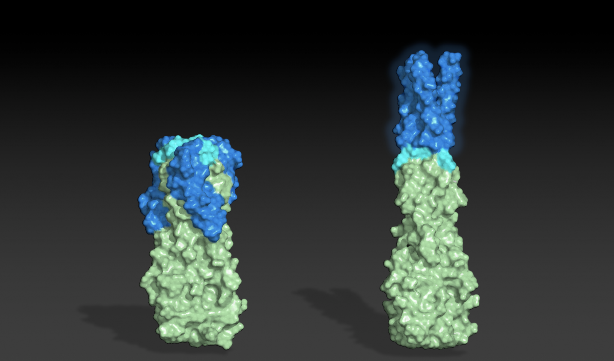
Computationally designed small-molecule sensors. Left: In the open state, ions flow freely through the transmembrane nanopore (cyan). Right: In the closed state, the sensor protein (pink) closes around the target small molecule, physically blocking the flow of ions and altering the membrane potential. A target molecule’s presence can be detected by measuring changes in conductance across the lipid membrane.
This research was led by Baker Lab researchers Samuel Berhanu, Sagardip Majumder, and Linna An. Scientists from the University of Basel, University of Leeds, University of Virginia, Lawrence Berkeley National Laboratory, VIB-VUB, and the Institute for Protein Design Core R&D Labs also contributed to the structural and functional analyses of the new proteins.
Sagardip Majumder, PhD
Samuel Berhanu, PhD
Linna An, PhD
The biosensors were created in stages and reported across two papers. Together, these projects yielded entirely synthetic receptors that resemble those in the nose. “This collaboration is a great example of what’s possible with protein design,” said senior author and IPD director David Baker. “Rather than repurposing biomolecules from nature, we can now create the functions we want from first principles.”
Sculpting new nanopores
In the first study, the researchers designed custom protein nanopores and characterized them in the lab. These tube-like molecules have openings, or pores, less than three nanometers wide — roughly 20,000 times smaller than the diameter of a human hair.
The team showed that these custom nanopores embed into lipid membranes, similar to nasal scent receptors, and function as conduits for electrically charged ions as intended.
“Natural protein nanopores are important tools for DNA sequencing, but only a handful of these delicate molecules are reliable enough to work with. For this project, we created more stable nanopores than anyone’s had access to, and there’s effectively no limit to the number we could make using this design approach,” said Samuel, now a research scientist at IEH Laboratories and Consulting Group.
“This work brings biology and electronics much closer together. We’re now exploring how to incorporate them into bio-electronic devices that can detect tiny traces of chemicals, diagnose diseases, and possibly form critical components of nanoscale filtration devices,” said Sagardip.
Binding to chemicals
The second study introduced a novel approach for creating proteins that bind to specific chemicals.
The team chose four challenging small molecules as binding targets. These included cholic acid, an important marker for liver disease; methotrexate, an anti-folate cancer treatment that requires regular blood monitoring; and thyroxine, a hormone used to diagnose thyroid conditions.
“It hasn’t been easy to detect chemicals like these, but we succeeded in generating proteins that recognize each of our four targets,” said Linna. “The same approach could be used to create sensors for virtually any small molecule.”
Using a variety of deep-learning tools, the team generated “pseudocycle” proteins. These consist of repeating structural units that surround central binding pockets of varying shapes. They then docked target chemicals into these pockets and optimized the interaction surfaces using LigandMPNN, a deep-learning tool expanded from ProteinMPNN. The highest affinity binding proteins were identified via laboratory screening.
Creating synthetic sensors
By combining the small molecule binders within the new nanopores, the team created proteins that change conductance in response to target molecules — essentially creating synthetic tools that act like olfactory receptors.
Linna believes that synthetic nanopore sensors are just one technology that will emerge from this research. “When I talk to other scientists, they’re excited by how these binding proteins may be used in many different detection systems,” she commented. In addition to the nanopore sensors, the team describes chemically induced dimerization (CID) systems that could be used to control cancer cell therapies and more.
This research opens the door to developing highly specific biosensors for detecting disease biomarkers, monitoring environmental pollutants, and advancing personal healthcare devices. The collaboration underscores the interdisciplinary nature of modern science and the potential for computational protein design to revolutionize how we create solutions to important challenges in medicine and beyond.
This research was supported by several federal, private, and philanthropic organizations. All funding sources are listed in the manuscripts.
Berhanu S, Majumder S, Müntener T, et al. Sculpting conducting nanopore size and shape through de novo protein design. Science, 18 July 2024
Ah L, et al. Binding and sensing diverse small molecules using shape-complementary pseudocycles. Science, 18 July 2024
Researchers from the Manchester Institute of Biotechnology (MIB) and the Institute for Protein Design (IPD) have launched an initiative to transform the landscape of enzyme design.
Established today, the International Centre for Enzyme Design (ICED) will bring together internationally leading research teams to establish a fully integrated computational and experimental platform to develop a new generation of industrial biocatalysts.
Led by professor Anthony Green PhD, interim director of the MIB, along with professor Nicholas Turner, PhD, and Sarah Lovelock, PhD, and in partnership with IPD director David Baker, PhD, ICED aims to deliver customized biocatalysts for sustainable production of a wide range of chemicals and biologics, including pharmaceuticals, agrochemicals, materials, commodity chemicals, and advanced synthetic fuels.
“I am truly excited to establish this International Center for Enzyme Design with our academic and industrial partners. The centre aims to develop a new generation of predictive enzyme design and engineering technologies that allow the rapid delivery of customised biocatalysts to meet diverse industry needs.”
Natural and engineered enzymes can be used to speed up important chemical processes. This technology is now widely recognized as a key enabler of a greener and more efficient chemical industry.
Although powerful, existing experimental methods for developing industrial biocatalysts are costly and time-consuming. This limits the benefits that biocatalysis may have on many industrial processes. Furthermore, for many desirable chemical transformations, there are no known enzymes that can serve as starting templates for experimental engineering.
“Accurately designing efficient enzymes with new catalytic functions is one of the grand challenges for the protein design field. We are thrilled to be working with Professor Green and his team in the MIB to address this crucial biotechnological challenge.”
— Professor David Baker, PhD
In ICED we will bring together leading computational and experimental teams from across academia and industry to bring about a step-change in the speed of biocatalyst development. The approaches developed will also allow for the creation of new families of enzymes with catalytic functions that are unknown in nature.
The design tools developed throughout the project will be made available to specialists and non-specialists to support their own enzyme engineering and biocatalysis needs. As the center develops, we expect to grow our partnerships with the wider academic and industrial sectors to ensure that we can best serve the needs and ambitions of the global biocatalysis community.
The White House announced this week that the Institute for Protein Design will be among the first organizations awarded access to the National AI Research Resource (NAIRR). This program aims to democratize advanced AI research by providing academics with large-scale computing resources.
“Computational biologists have never had a way to get access to [computing] at this level,” said IPD director David Baker, PhD, in an interview with Science. “It’s hard for academics to keep up with industry.”
Rohith Krishna, a Baker Lab graduate student, attended the May 6 White House event where all 35 initial NAIRR pilot projects were announced. These projects span from modeling the geological effects of climate change to developing AI systems that can proactively identify deepfakes.
“NAIRR support will allow us to develop the next generation of protein structure prediction and design algorithms which will have applications in human health and sustainability,” explains Krishna, who presented a brief overview of this work at the White House.
Rohith Krishna at the White House on May 6, 2024. Image: Charlotte Geary/NSF
Rohith Krishna presenting at the White House on May 6, 2024. Image: Charlotte Geary/NSFRohith Krishna presenting at the White House on May 6, 2024. Image: Charlotte Geary/NSFImage: Charlotte Geary/NSFArati Prabhakar, PhD, Director of the White House Office of Science and Technology Policy (OSTP) and Assistant to the President for Science and Technology. Image: Charlotte Geary/NSF
NAIRR was created as part of President Joe Biden’s October 2023 executive order on AI. He directed the the National Science Foundation (NSF) to lead the program which draws resources, at least initially, from supercomputing facilities supported by NSF and the Department of Energy. Our pilot project, Advanced Training for Protein Diffusion, Binder Prediction, and Antibody Design, will be supported until October 2024 by theTexas Advanced Computing Center at The University of Texas at Austin.
With NAIRR support, our scientists are poised to make even greater strides at the intersection of AI and protein science. We intend to continue our traditions of responsible and open science, having hosted the world’s first AI safety summit in our field and having released our most powerful AI systems for protein modeling and design as free and open tools that all scientists may use.
The organizations receiving pilot NAIRR support span 17 states and will include national research centers, the Mayo Clinic, and over 20 universities. The University of Washington was issued three pilot awards, more than any other organization.
About the Institute for Protein Design
Located at one of the largest and most innovative public universities, the Institute for Protein Design at the University of Washington School of Medicine is a world-renowned center for computational biology. Our mission is to develop advanced tools for protein science and use them to solve modern challenges in medicine, technology, and sustainability. Our researchers are pioneering AI tools for a wide range of applications, including drug discovery, vaccine design, and materials science.
David Baker, PhD, is the director of the Institute for Protein Design, an HHMI Investigator, and a professor of biochemistry at the University of Washington School of Medicine. For decades, his lab has developed state-of-the-art protein design software and used it to create molecules that solve challenges in medicine, technology, and sustainability. Among his recent work is the development of powerful AI tools for generating functional proteins.
David’s achievements include publishing over 600 peer-reviewed papers, training over 80 professors, co-founding 21 biotechnology companies, and securing over 100 patents. A recipient of the Breakthrough Prize, his work was recognized in 2021 by the journal Science as the most significant across all domains of research.
TIME includes David Baker among its list of Health Pioneers, writing:
From TIME:
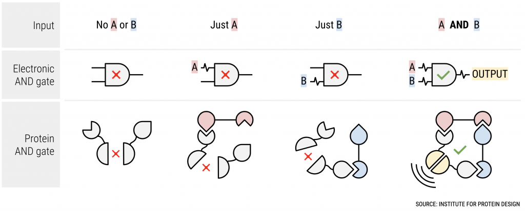
Much of Baker’s early research was aimed at understanding how proteins fold. But in the 1990s, after developing a software program, Rosetta, to help answer this question, Baker and his research team realized that they could, in essence, run the software backwards, and design a protein based on a desired shape. In recent years, Baker and researchers in his lab have designed proteins that act as biological “logic gates,” allowing scientists to program cellular functions, such as gene expression, just as they would a computer, and a protein-based antiviral nasal spray that targets COVID-19.
Baker has co-founded 17 companies and been granted over 100 patents. Rosetta has evolved into RosettaCommons, one of the most widely-used protein design software packages. For his research, Baker, now director of the Institute for Protein Design at the University of Washington, was awarded the 2021 Breakthrough Prize in Life Sciences.
Recently, Baker has been a leader in grappling with the societal implications of the technologies he has helped create. Rapid advances in artificial intelligence have sparked fears that AI systems could exacerbate risks of bioterrorism. In response, Baker shepherded an agreement this year, signed by more than 90 scientists in the field, that commits to promoting the responsible development and use of AI protein-design tools.
Read the full article by Will Henshall at Time.com
Two of our undergraduate researchers — Abby and Sneha — have been recognized by the University of Washington as leaders and innovators.
The Husky 100 includes undergraduate, graduate, and professional students who have founded startups, created artwork, served as mentors, conducted research, and advocated for social justice. In honor of their many contributions, each is eligible to participate in a range of activities and opportunities offered by our on- and off-campus partners.
Congrats Abby and Sneha!
Abby Burtner
From: Olympia, WA
B.S. Biochemistry, Chemistry and Data Science
“Through my academic, research and community-building experiences at the UW, I have discovered that I want to pursue a career in science. I aspire to be a professor leading a research team investigating the molecular mechanisms of the immune system. I hope to harness this knowledge to design therapeutics for globally impactful infectious diseases and underresearched autoimmune diseases.”
Sneha Subramanian
From: Bellevue, WA
B.S. Public Health-Global Health
“Witnessing pervasive health disparity, I am driven to pursue research, leadership, and community initiatives that promote accessibility in medicine. The University of Washington has equipped me with indispensable experiences, enabling me to harness public health frameworks to dismantle institutional barriers that perpetuate inequity in healthcare. I am dedicated to leveraging my unique lived experiences to catalyze systemic change within health systems, while providing empathetic and culturally competent care to diverse populations worldwide.”
About the Husky 100
Each year, the Husky 100 honors the outstanding work and achievements of 100 students on all three UW campuses who are making the most of their Husky Experience.
IPD Executive Director Lynda Stuart, MD, PhD, recently spoke at the Life Science Innovation Northwest conference. She was joined by panelist from Absci, Bristol Myers Squibb, Microsoft, NVIDIA.
From GeekWire:
“The Pacific Northwest has the compute, it has the biotech, but it also has a kind of culture of collaboration and sharing that is not present in certain other parts of the country,” said Lynda Stuart, executive director of the Institute for Protein Design (IPD) at the University of Washington.
“A regional hub is a very natural thing to emerge,” said Portland, Ore.-based Jonathan Cohen, vice president of applied research at NVIDIA, which is investing heavily in AI-mediated drug design.
The IPD is at the center of this hub. The IPD generates open-source AI tools to craft protein-based therapeutics, vaccines, materials and biosensors, and its Seattle-area spinouts and affiliated companies interact with each other and partner with larger biopharma companies.
In September, Redmond, Wash.-based Microsoft released an open-source model to generate proteins, and other big tech companies are also betting on the field.
One major aim is to not only discover new therapeutic proteins but to shorten their clinical development through “quality by design,” said Stuart. Researchers can now assess proteins for traits such as ease of manufacture or unwanted cross-reactivity to other molecules, she said.
Read the full story by Charlotte Schubert at geekwire.com
Scientists in the Baker Lab published a preprint in March showing that RFdiffusion can be tuned to generate antibodies. Laboratory testing confirmed that these proteins can bind the influenza virus and other targets as intended.
This proof-of-concept study was recently covered by Nature.
From Nature:
Antibodies — immune molecules that strongly attach to proteins implicated in disease — have conventionally been made using brute-force approaches that involve immunizing animals or screening vast numbers of molecules.
AI tools that can shortcut those costly efforts have the potential to “democratize the ability to design antibodies”, says study co-author Nathaniel Bennett, a computational biochemist at the University of Washington in Seattle. “Ten years from now, this is how we’re going to be designing antibodies.”
“It’s a really promising piece of research” that represents an important step in applying AI protein-design tools to making new antibodies, says Charlotte Deane, an immuno-informatician at the University of Oxford, UK.
Read the full story by Ewen Callaway at nature.com
What do squirrel skeletons and fish faces have in common with vaccines and bat flight? Probably only one thing: undergraduate researcher Abby Burtner has studied them all.
Already named a Goldwater Scholar, Mary Gates Scholar, and Washington Research Foundation Fellow, Abby has been conducting research since her freshman year. We’re delighted to share that she has also just been named a 2024 Churchill Scholar in recognition of her outstanding achievements in the field of biochemistry.
Abby attributes her success in science to all those who have supported her so far, including multiple professors, labmates, family members, and more. Photo by Jayden Becles
“The Churchill scholarship,” says Ed Taylor, vice provost and dean of Undergraduate Academic Affairs, “is a prestigious opportunity for Abby to continue expanding her biochemistry skills. This award reflects her capacity to draw from her research and the mentorship she’s experienced, to fuel her work toward a greater understanding of our world in critical ways. The UW’s research community and campus-at-large are proud of Abby and encourage her as she continues to live out UW’s mission at Cambridge.”
Beginning in 2022, Abby has conducted protein design research in the King Lab, which is part of the Institute for Protein Design. Her work here has focused on sculpting how the immune system responds to vaccines, with the goal of creating “sidekicks,” or adjuvants, that will help unlock durable protection following vaccination.
Abby’s curiosity for biology — nurtured during childhood rafting trips in the Pacific Northwest and honed through research she’s conducted here and abroad — was recently profiled by the University of Washington.
Congratulations, Abby!
From UW Undergraduate Academic Affairs:
Broadened perspectives
An interdisciplinary history and philosophy class helped shape Burtner’s perspective on science and innovation. The class’s exploration of scientific revolutions sparked her realization that advancements in one field can revolutionize another. “That was an interesting look at the science that made me take a step back,” she said of the genesis of her interdisciplinary studies. This cross-disciplinary awakening to new ideas and connections guided Burtner directly to Professor Neil King’s lab at the Institute for Protein Design (IPD).
Burtner’s arrival at the lab coincided with South Korea approving the IPD’s COVID-19 vaccine. Burtner was now working at the forefront of much-needed vaccine innovations. The IPD has revolutionized medicine and protein design through deep learning, a method in artificial intelligence (AI) that teaches computers to process data like the human brain, and machine learning, the development of statistical algorithms that learn from known data and unseen data. “When you are not limited by technology anymore, it comes down to how creative you can be,” Burtner explained.
Changing the world
“I think vaccines are particularly dramatic examples of technologies that can change the world for the better.”
Abby Burtner
Many traditional vaccines, like the flu vaccine, use a virus that’s live, but weakened or dead to stimulate strong immune responses. “There are huge public health ramifications,” said Burtner, explaining that these processes can cause issues for immunocompromised individuals. Research is shifting toward safer protein-based subunit vaccines, which eliminate potential risks by only displaying the necessary components of the pathogen.
“When people in my life have become sick, I want to help them,” said Burtner of her drive to help others through research. These vaccines required the co-delivery of adjuvants, like aluminum salt, to stimulate the immune system, but their mechanism isn’t fully understood and can’t be tailored for a specific immune response. Burtner sought out to create protein-based adjuvants to co-deliver with protein vaccines for a safer, more effective approach.
By targeting the toll-like receptor family of proteins, well-known activators of immune signaling, the goal was to get those specific immune responses.
“The IPD has been an inspiring space to be because of rapid design developments worldwide,” said Burtner. She cites the lab’s innovative and revolutionary design of de novo proteins as possible due to the deep learning revolution in biochemistry. The breakthrough release of AlphaFold2, the protein structure prediction algorithm enabled them for the first time to accurately predict the structure of a protein.
IPD director David Baker recently spoke at START SOMETHING, a series by UW CoMotion that features conversations with entrepreneurs and innovators.
This post highlights key points from the event, moderated by Jenny Cronin, principal at the Allen Institute for AI (AI2) Incubator. The two spoke about the impact of artificial intelligence on life sciences and healthcare startups. Their full conversation is available on YouTube.
Integrating AI into Protein Science
The Institute for Protein Design is at the center of a technological revolution. For decades, David’s group and others developed and used traditional physics-based software such as Rosetta to model and design biomolecules. Today, AI-driven approaches are transforming this work.
Where does change like this come from? “We have a lot of people wanting to collaborate and visit, and they often bring in ideas,” David said.
AI tools for protein design have enhanced our ability to create molecules with advanced functions, including experimental vaccines, medicines, nanomaterials, and much more. Many of the most powerful and popular AI-based tools for protein science were created here at UW Medicine.
AI’s Impact on Therapeutics and Beyond
From shortening the time it takes to respond to new viruses to enhancing treatments for solid tumors, David sees profound medical potential in these tools. “I think we’re going to see a transition from protein therapeutics being obtained by kind of black magic — immunizing an animal and letting the natural immune system come up with a solution — to actually being designed rationally,” he said.
Beyond delivering the world’s first computer-generated protein medicine, scientists here at the Institute for Protein Design are advancing research in non-therapeutic areas. This includes developing artificial photosynthetic systems and custom nanopores for environmental monitoring.
We have elected to make our most powerful AI-driven tools for protein design, including RFdiffusion and ProteinMPNN, completely open source, meaning any researcher in the world can use them or improve on them at no cost. David discussed how this strategy has allowed for much greater collaboration and innovation, leading to a worldwide explosion in new protein design technologies and applications.
David’s advice for budding scientists and entrepreneurs: be open to collaborations and reach out to experts in your field. “I encourage my students to just email people. If they’re working on a problem, I say figure out who the best three people are in the world and email them. Sometimes you won’t get an answer, but other times you will, and that can start a connection which could really transform your research.”
The Commercial Potential of Scientific Research
Our Institute fosters entrepreneurship in many ways, including through our Translational Investigator Program and by encouraging open dialogue among researchers within and beyond our walls.
“If you choose the right problems, where this is the time to solve them, a surprisingly large fraction will have some commercial potential — maybe in a way that you didn’t anticipate,” David explained. “That certainly happened with a lot of the things we’ve done.”
David’s commitment to his homecity Seattle has contributed to the city becoming a thriving ecosystem for biotech startups. Many of our Institute’s spinouts were formed and remain in Seattle, which has been pivotal in building this ecosystem. We look forward to launching many more companies in this vibrant city.
To maximize the benefits and minimizing risks of AI for protein design, IPD director David Baker and scores of other senior scientists across 20 countries have signed a community agreement with ten actionable commitments and invited others in the field to join them.
The Institute for Protein Design is playing a key role in an international initiative focused on the safe, secure, and beneficial development of AI tools for biomolecular research. Central to this is a voluntary community statement published today and signed initially by over 90 preeminent researchers in the field.
Leading the signatories is David Baker, PhD, director of the Institute for Protein Design and professor of biochemistry at UW Medicine. His lab’s contributions to AI-driven protein science were recognized by the journal Science as the 2021 Breakthrough of the Year. IPD colleague and UW assistant professor Neil King, PhD, also helped deliver the world’s first medicine produced through computational protein design.
“I view this as a crucial step for the scientific community. The responsible use of AI for protein design will unlock new vaccines, medicines, and sustainable materials that benefit the world. As scientists, we must ensure this happens while also minimizing the chance that our tools could ever be misused to cause harm.”
— David Baker, PhD
Scores of senior researchers working in over 20 countries have also signed the agreement, including 2018 Nobel laureate and Caltech professor Frances Arnold, PhD, and Microsoft’s Chief Scientific Officer, Eric Horvitz, MD, PhD. Both serve on President Biden’s Council of Advisors on Science and Technology, with Arnold as an external co-chair.
An open call for responsible AI
The community agreement, signed by these scientists in their personal capacities, included ten specific commitments to foster responsible AI innovation in the life sciences. These include thorough safety reviews of emerging AI models for protein design and applying these technologies to help create vaccines and medicines for global health challenges.
The scientists also call for improved security measures around DNA manufacturing, a critical step in the research pipeline where potential hazards could emerge. David and Harvard geneticist George Church, PhD recently proposed new DNA synthesis policy in the journal Science.
Today, all senior scientists in this field are invited to sign this agreement. Future gatherings are expected and will explore comprehensive implementation strategies, uniting scientists, security experts, and policymakers. The agreement was drafted with extensive input from scientists and informed by discussions with biosecurity and policy experts and professionals in other AI fields.
“We at the IPD are aware of the power of the new AI biodesign tools. It is fantastic to see this community come together in this way to ensures that they are used responsibly and for the advancement of all.”
— IPD Executive Director Lynda Stuart, MD, PhD
In addition to accepting signatures from scientists, the statement also lists a growing number of supporters who endorse the spirit of this effort. These include biosecurity professionals at the Coalition for Epidemic Preparedness Innovations and members of the Africa Centers for Disease Control and Prevention and the Council on Foreign Relations.
All signatories and supporters are listed at the community statement’s website, responsiblebiodesign.ai.
This initiative builds upon discussions at the AI safety summit we convened last October, emphasizing the importance of responsible management of AI technology and the roles scientific experts can play in shaping future policies. It exemplifies a collaborative spirit among individual scientists worldwide, showcasing their commitment to advancing technology for global benefit.
About the Institute for Protein Design
The Institute for Protein Design at the University of Washington School of Medicine is a global leader in science. From creating AI technologies to delivering the world’s first computationally designed protein medicine, our work reflects our dual commitment to both generate knowledge and achieve impact. Our mission is to create new proteins that solve modern challenges in medicine, technology, and sustainability.
The Institute for Protein Design exists to create scientific breakthroughs and transform them into practical solutions. Of the roughly 200 researchers who train or work here, about one third are women. This includes staff scientists, acting instructors, postdoctoral scholars, and undergraduate and graduate student researchers.
Unfortunately, these numbers are in line with science as a whole. According to the UN, women hold only one third of all research roles. They tend to have shorter, less well-paid careers and are often passed over for promotions. There are some research disciplines in which women make up the majority of working professionals, but in fast-moving fields such as artificial intelligence, women find themselves outnumbered by a factor of four.
As a scientist, physician, and black woman myself, I know what it’s like to feel alone in a crowded room. I also know the value of continuous support and the awesome power that examples can have. As Executive Director of this Institute, I will be making diversity, equity, and inclusion a priority. There is much more we can do to attract and retain women at all career stages, and that work has already begun.
Every one of our scientists helps make this place the research powerhouse that it is. Our women trainees and staff have made pioneering contributions to computer science, biochemistry, immunology, and more. Each embodies the relentless spirit of excellence that drives all of us at the Institute for Protein Design.
Here are three examples.
Image: Seoul National University
AI Pioneer
Minkyung Baek, PhD
Assistant Professor, Seoul National University
Minkyung has already achieved what most scientists only dream of: As a postdoctoral scholar in the Baker Lab, she completed a research project that helped transform an entire field of science. She led the development of RoseTTAFold, a powerful machine learning tool that accurately models the intricate shapes of proteins. That tool — along with AlphaFold, a similar technology by Google DeepMind — was recognized by the journal Science as the 2021 Breakthrough of the Year.
Following her time with us, Minkyung took up an assistant professorship at her alma mater. She now leads the Laboratory of Computational Structural and Systems Biology at Seoul National University. Minkyung is continuing to collaborate with researchers in the Baker and DiMaio Labs on new generations of RoseTTAFold, including a version that has been trained to model how DNA, RNA, and proteins interact.
“I do not believe that AI will replace humans. AI will more likely assist humans with what they need to do.”
— Minkyung Baek, PhD
Vaccine Innovator
Grace Hendricks
PhD Student, King Lab
Growing up in the America’s Southwest, Grace spent quite a few Saturdays at her mother’s workplace surrounded by walls full of chemicals and complicated posters. She recalls trying to entertain herself as her mom, a pharmacist, dispensed medicines.
Grace is paying a lot more attention to medicines these days, particularly vaccines. “During the COVID-19 pandemic, a lot of new technologies came online, including mRNA vaccines,” she explains. “The Institute for Protein Design also released a coronavirus vaccine based on computationally designed proteins.” As a graduate student in the King Lab, Grace is now working to combine these two technologies to create vaccines that are more potent and rapidly manufacturable. “We just want to make the best vaccines we can using all of the technology that’s available to us.”
Nowadays, Grace talks to her mother quite often about medicines. She shares updates about her own vaccine research and even answers questions about the updated COVID-19 booster shots. During these conversations, Grace sometimes reflects on those days spent at the pharmacy with her mother.
“If my mom hadn’t had to take me with her into work on the weekends, maybe I wouldn’t have seen all those posters and all those medicines on the wall. And I sometimes wonder, would I still be here today?”
— Grace Hendricks
Anti-venom aficionado
Susana Vázquez Torres
PhD Student, Baker Lab
In December, Susana and colleagues published in Nature a new approach for generating proteins that bind to target molecules with remarkable affinity. She believes this AI-enabled advance is poised to “redefine the landscape of biotechnology.”
Susana appeared on NPR’s Morning Edition to share how she is using this technology to develop medicines that shut down snake venom. In an interview with NPR science reporter Geoff Brumfiel conducted in the Baker Lab, her excitement around emerging technologies that can be used to improve lives was clear to hear.
To maintain biosecurity in the age of AI, all synthesized DNA sequences should be screened and logged, according to two leading scientists. Such records could be decrypted and scrutinized in the event of a novel biological threat, a practice that may help deter the misuse of biodesign software.
David Baker and George Church — two professors whose research has helped define modern biotechnology — published an editorial in Science this week calling for enhanced screening and universal logging for all manufactured DNA sequences.
“This would present a practical barrier to the creation of harmful biomolecules, whether accidental or intentional,” they write. Their commentary, Protein design meets biosecurity, was published Jan 25.
From Science
“The power and accuracy of computational protein design have been increasing rapidly with the incorporation of artificial intelligence (AI) approaches. This promises to transform biotechnology, enabling advances across sustainability and medicine. DNA synthesis plays a critical role in materializing designed proteins. However, as with all major revolutionary changes, this technology is vulnerable to misuse and the production of dangerous biological agents. To enable the full benefits of this revolution while mitigating risks that may emerge, all synthetic gene sequence and synthesis data should be collected and stored in repositories that are only queried in emergencies to ensure that protein design proceeds in a safe, secure, and trustworthy manner.”
David Baker, PhD, is the director of the Institute for Protein Design and a professor of biochemistry at the University of Washington. His research group develops and uses software for protein design. In recent years, they have created powerful deep-learning technologies, including RFdiffusion and ProteinMPNN, that can be used to generate biomolecules with new functions.
George Church, PhD is the Robert Winthrop professor of genetics at Harvard Medical School and a leading figure in the development of modern DNA sequencing and synthesis technologies. He has been involved in creating biosecurity policy for over 20 years. He is also a member of our Advisory Board.
Our commitment to AI safety and security
Last October, we convened a first-of-its-kind summit on the responsible development of AI technologies in the field of protein design. Our event brought together leading academics from around the world and representatives from other sectors including private industry, non-profits, and the US and UK governments. Security concerns and solutions were discussed.
The editorial published by David and George this week was inspired by conversations that occurred at our summit. The need for community guidelines to drive the development of safe and secure AI technologies was also emphasized at the event. Those guidelines are in development and will be shared soon.
AI-enabled protein design is a technology area you’ll want to keep an eye on, according to Nature. With massive training datasets and ever more sophisticated deep-learning approaches, tools like our own RFdiffusion All-Atom are opening the door to custom enzymes, advanced biomaterials, and more.
From Nature:
Deep learning for protein design
Two decades ago, David Baker at the University of Washington in Seattle and his colleagues achieved a landmark feat: they used computational tools to design an entirely new protein from scratch. ‘Top7’ folded as predicted, but it was inert: it performed no meaningful biological functions. Today, de novo protein design has matured into a practical tool for generating made-to-order enzymes and other proteins. “It’s hugely empowering,” says Neil King, a biochemist at the University of Washington who collaborates with Baker’s team to design protein-based vaccines and vehicles for drug delivery. “Things that were impossible a year and a half ago — now you just do it.”
Much of that progress comes down to increasingly massive data sets that link protein sequence to structure. But sophisticated methods of deep learning, a form of artificial intelligence (AI), have also been essential.
‘Sequence based’ strategies use the large language models (LLMs) that power tools such as the chatbot ChatGPT (see ‘ChatGPT? Maybe next year’). By treating protein sequences like documents comprising polypeptide ‘words’, these algorithms can discern the patterns that underlie the architectural playbook of real-world proteins. “They really learn the hidden grammar,” says Noelia Ferruz, a protein biochemist at the Molecular Biology Institute of Barcelona, Spain. In 2022, her team developed an algorithm called ProtGPT2 that consistently comes up with synthetic proteins that fold stably when produced in the laboratory [1]. Another tool co-developed by Ferruz, called ZymCTRL, draws on sequence and functional data to design members of naturally occurring enzyme families [2].
Sequence-based approaches can build on and adapt existing protein features to form new frameworks, but they’re less effective for the bespoke design of structural elements or features, such as the ability to bind specific targets in a predictable fashion. ‘Structure based’ approaches are better for this, and 2023 saw notable progress in this type of protein-design algorithm, too. Some of the most sophisticated of these use ‘diffusion’ models, which also underlie image-generating tools such as DALL-E. These algorithms are initially trained to remove computer-generated noise from large numbers of real structures; by learning to discriminate realistic structural elements from noise, they gain the ability to form biologically plausible, user-defined structures.
RFdiffusion software [3] developed by Baker’s lab and the Chroma tool by Generate Biomedicines in Somerville, Massachusetts [4], exploit this strategy to remarkable effect. For example, Baker’s team is using RFdiffusion to engineer novel proteins that can form snug interfaces with targets of interest, yielding designs that “just conform perfectly to the surface,” Baker says. A newer ‘all atom’ iteration of RFdiffusion [5] allows designers to computationally shape proteins around non-protein targets such as DNA, small molecules and even metal ions. The resulting versatility opens new horizons for engineered enzymes, transcriptional regulators, functional biomaterials and more.
Read the full article by Michael Eisenstein at nature.com
The article features an illustration (shown above) by The Project Twins.
With a $100,000 Phase 1 commercialization grant from the Washington Research Foundation, IPD researchers Alexis Courbet, PhD, and Jinwei Xu, PhD, are aiming to create the first direct interface between biochemistry and electronics for multi-omics applications.
This project leverages AI-based protein design to create custom protein nanopores that can be integrated within semiconductors, setting the stage for a new era in nanopore technology. It began as fundamental research on protein nanopore design here at the Institute for Protein Design.
“By bridging biology and electronics, we’re creating a new way to extract vast amounts of data from biology. This could be transformative for precision medicine, allowing for a greater understanding of human health and new approaches for treating disease faster and closer to patients,” explains Courbet.
“Beyond medicine, we also believe this technology will make it easier and cheaper to interface with the environment in a highly multiplexed way, leading to better detection of contaminants and other important substances,” adds Xu.
A New Era in Biosensing
Biosensors — which are used to detect DNA, antibodies, and other chemicals — are becoming indispensable in healthcare, environmental monitoring, and forensics. In medicine, an estimated 70 percent of clinical decisions are now underpinned by some form of diagnostic technology. Our project aims to create biosensor technology unlike any available today, with the goal of enabling more holistic and accurate disease modeling, biomarker discovery and detection, and environmental monitoring.
“While some natural protein nanopores are repurposed today primarily for DNA sequencing, their delicate structure and complex biochemistry make them challenging to work with,” notes Xu. “Recent breakthroughs in AI-enabled protein design now make it possible to create nanopores from scratch that do exactly what we want them to do. We’re hoping this will unlock a new era of nanopore technologies with utilities far beyond DNA sequencing.”
Fundamental research conducted at our Institute has shown that novel protein nanopores can be created through protein design and that these molecules can be much more robust and amenable to downstream modifications than their natural counterparts.
Biotechnology Powered By Generative AI
At the core of this project is the pioneering use of generative AI for protein design, allowing for the systematic creation and integration of custom protein nanopores directly within semiconductors. When combined with state-of-the-art nanolithographic manufacturing processes, this leads to the formation of protein-silicon devices capable of achieving extremely high nanopore sensor densities.
Courbet and Xu estimate that they can create devices with approximately one million times more protein sensors than are found in today’s commercial protein-based biosensor technologies. Such extreme densities may offer many advantages, including enhanced detection of rare compounds and significantly enhanced multiplexing, promising to redefine the landscape of multi-omic biomarker detection for patients in a healthcare setting and to expand sensitive multianalyte detection in environmental contexts.
“Compared to lipid-based nanopore approaches, we hope to greatly enhance the volume of multidimensional data being generated by our biosensing platform and to seamlessly transfer these data through bioelectronic devices to enable on-device information processing,” explains Courbet.
Milestones and Additional Support
The WRF-supported milestones for this project are ambitious. They include designing and characterizing protein nanopore adaptors, achieving high-resolution DNA sequencing, and developing stable electrokinetic docking of proteins on solid-state nanopores, among others.
This initial commercialization grant complements a $1.5 million discovery research grant already received from the Bill and Melinda Gates Foundation, underscoring the broad support and potential of this work. Our fundamental research on protein nanopore design has also been supported by The Audacious Project and other funders.
This advance could allow scientists to create cheaper alternatives to antibodies for disease detection and treatment.
This week we report in Nature an AI-enabled advance in biotechnology with implications for drug development, disease detection, and environmental monitoring. Using a combination of traditional and deep learning based molecular design approaches, we’ve created proteins that bind with exceptionally high affinity and specificity to a variety of challenging biomarkers, including human hormones. Notably, we achieved what we believe to be the highest binding affinity ever reported between a computer-generated biomolecule and its target.
AI-enabled protein design software in action. Beginning with a desired binding target (pink) and a cloud of disconnected amino acids, RFdiffusion iteratively sculpts a new protein structure that cradles the target peptide. At the end, ProteinMPNN assigns amino acid side chains to the new protein structure, yielding a complete protein molecule. Laboratory tests reveal this protein binds its target with the highest affinity ever reported to date for a computer-generated protein without any experimental optimization.
This project was led by Baker Lab members Susana Vazquez-Torres, Preetham Venkatesh, and Phil Leung, PhD. It included collaborators from UW Medicine and the University of Copenhagen, as well as from our Core R&D Labs.
The team set out to create proteins that could bind to glucagon, neuropeptide Y, parathyroid hormone, and other helical peptide targets. Such molecules, crucial in biological systems, are especially challenging for drugs and diagnostic tools to recognize as they often lack stable molecular structures. Antibodies can be used to detect some of these targets but are often costly to produce and have limited shelf lives.
“There are many diseases that are difficult to treat today simply because it is so challenging to detect certain molecules in the body. As tools for diagnosis, designed proteins may offer a more cost-effective alternative to antibodies,” explains Venkatesh.
The role of generative AI
The study introduces a novel way of using RFdiffusion, a generative model for creating new protein shapes, in conjunction with the sequence-design tool ProteinMPNN. Developed in the Baker Lab, these programs allow scientists to create functional proteins more efficiently than ever before. By combining these tools in new ways, the team was able to create binding proteins by using limited target information, such as a peptide’s amino acid sequence alone.
“We’re witnessing an exciting era in protein design, where advanced artificial intelligence tools, like the ones featured in our study, are accelerating the improvement of protein activity. This breakthrough is set to redefine the landscape of biotechnology,” notes Vazquez-Torres.
Measuring binding in the lab
In collaboration with the Joseph Rogers Lab at the University of Copenhagen and Andrew Hoofnagle Lab at UW Medicine, we conducted laboratory tests to validate the new biodesign methods.
Mass spectrometry was used to detect designed proteins that bind to low-concentration peptides in human serum, thereby demonstrating the potential for sensitive and accurate disease diagnostics. Additionally, the proteins were found to retain their target binding abilities despite harsh conditions including high heat, a crucial attribute for real-world application.
From binders to biosensors
With improved methods for creating binding proteins in place, the team turned to the challenge of designing new biosensors. To make sensors that could detect parathyroid hormone (PTH), we grafted a high-affinity PTH binder into our previously reported lucCage biosensor system. The best-performing biosensor lit up when mixed with PTH, displaying a 21-fold increase in bioluminescence.
“The ability to design proteins with such high affinity and specificity opens up a world of possibilities, from new disease treatments to advanced diagnostics,” concludes senior author and Institute director David Baker.
Funding
This work was supported by the National Institutes of Health (T1D U01 DK121289, U19 AG065156, K99EB031913, P30 GM124165), National Science Foundation (EF-2021552), Department of Energy (BER-ERCAP0022018; DE-AC02-06CH11357), European Molecular Biology Organization (ALTF 292-2022), Washington State General Operating Fund, Amgen, Audacious Project, AWS, Bill and Melinda Gates Foundation (INV-010680), Donald and Jo Anne Petersen, Howard Hughes Medical Institute, Microsoft, Novo Nordisk Foundation (NNF19OC0054441), Open Philanthropy Project, and Partnership for Clean Competition.
Today we published new research that may one day be applied to help remove large amounts of excess carbon from the environment.
In a paper appearing in Nature Communications, we show that custom proteins can drive the formation of carbon-rich minerals in laboratory settings. This offers a potential pathway for enhanced carbon storage via engineered organisms. This research was performed in collaboration with the Center for the Science of Synthesis Across Scales (CSSAS), which is co-led by the UW and Pacific Northwest National Laboratory (PNNL).
“In nature, proteins are behind the growth of bones, shells, and other durable materials like limestone. We’re now exploring how custom proteins can be used to create a wide range of new, robust materials. This study is an important first step, showing we can guide mineral formation in novel ways,” explains senior author David Baker.
The project was led by Baker Lab members Fatima Davila-Hernandez and Harley Pyles, and PNNL postdoctoral research associate Biao Jin of the De Yoreo research group. Jim De Yoreo is a Battelle Fellow at PNNL, an affiliate professor of materials science and engineering and of chemistry at the UW, and the Deputy Director of CSSAS.
The team used advanced molecular design software to create proteins with custom lengths and surface chemistries. They also showed that these designed features can influence the formation and growth of calcium carbonate crystals, a key component of seashells and limestone. The process by which biomolecules such as proteins promote mineral growth is called biomineralization.
With the aid of powerful microscopes, tiny crystals of protein-stabilized calcium carbonate can be seen bunching together.
“By combining different imaging techniques, I could see the tiny crystals form around our proteins and grow bigger by the second.”
Baio Jin, PhD, co-lead author
Biology’s Role in Earth’s Carbon Cycle
Our research paves the way for future studies on calcium carbonate formation under biological conditions, which is a critical part of the global carbon cycle. This line of research could also help in the fight against climate change by offering new routes to large-scale and long-term carbon sequestration.
“Our designed proteins are an important step toward mimicking nature’s efficiency in carbon storage. While we’re not yet at nature’s level, we are gaining valuable insights that may inform the development of practical, scalable solutions for long-term carbon storage,” said Pyles.
Whether in human teeth or aquatic microbes, nature’s mineral-forming proteins contain irregular features that make them difficult for scientists to study, even with advanced instruments or the latest protein modeling tools like RoseTTAFold All-Atom. By designing new mineral-forming proteins from scratch, we hope to enable controlled research that will reveal the interplay between the living and nonliving worlds.
Addressing Climate Change
Combatting human-caused climate change will require many innovative solutions, including finding effective ways to remove carbon pollution from the environment. Organisms like kelp, algae, and trees naturally store carbon atoms in their tissues through photosynthesis, but their capacity for long-term storage is limited. In contrast, some aquatic microorganisms convert carbon in ocean water into calcium carbonate crystals using biomineralization, leading to sedimentation and limestone formation. However, this natural process is slow.
Integrating designed proteins into living organisms may one day accelerate limestone formation in the ocean, potentially transforming billions of tons of carbon pollution into enduring mineral deposits. Rigorous evaluations of the feasibility and environmental impacts of this research will be needed before any real-world application.
Keep an eye out for more updates from us and our colleagues as we continue to explore the potential of protein design to create solutions across medicine, technology, and sustainability.
Join Us
Are you intrigued by the prospect of using protein design to solve modern challenges? We invite you to join us or support our work.
Funding
This work was supported by numerous funding sources, including the United States Department of Energy Office of Science and The Audacious Project at the Institute for Protein Design. A full list of funders is provided in the paper.
Today we learned that Icosavax, a Seattle-based vaccine design company born from innovative research conducted here at the Institute for Protein Design, will be acquired by AstraZeneca in a deal worth up to $1.1 billion.
“This is a significant moment for the UW School of Medicine, showcasing how fundamental research can lead to groundbreaking technologies that stand to benefit the world,” said Tim Dellit, CEO of UW Medicine, the Paul G. Ramsey Endowed Dean of the UW School of Medicine and the university’s executive vice president for medical affairs.
According to the UK-based drug maker, the planned acquisition will strengthen AstraZeneca’s late-stage vaccine development pipeline, with Icosavax’s lead vaccine candidate, IVX-A12, now poised to become the first-in-class, Phase III-ready, combination vaccine targeting two major respiratory viruses that burden older adults and those with certain chronic illnesses.
A Breakthrough in Vaccine Development
Launched in 2017 with Neil King and David Baker as scientific co-founders, Icosavax was formed in our Translational Investigator Program with support from the UW innovation office CoMotion. The company’s approach, based on computationally designed self-assembling protein nanoparticle technology developed here, leverages significant new capabilities in how vaccines can be constructed at the molecular level.
“We’re thrilled that vaccine technology developed at our public university may soon protect many people from respiratory disease.”
Neil King, head of vaccine design at the Institute for Protein Design and assistant professor of biochemistry at UW Medicine
King led early research on designed protein self-assembly as a postdoctoral scholar in the Baker Lab and helped this technology platform mature as an IPD Translational Investigator.
David Baker, PhD, (left) and Neil King, PhD, (right) both holding 3D-printed models of designed protein nanoparticles.
Protein nanoparticles mimic the round shape of natural viruses and can be decorated with key viral fragments, yielding custom vaccine components capable of triggering robust protective immune responses. This novel vaccine science not only promises to enhance efficacy but may also minimize some of the side effects of vaccination, as seen by ongoing research from the King Lab.
Our protein nanoparticle technology used by Icosavax has already produced a licensed vaccine product. The COVID-19 vaccine SKYCovione, developed by the King and Veesler Labs at UW Medicine and advanced into the clinic by our collaborators at SK Bioscience, is the world’s first computationally designed protein-based medicine.
Icosavax’s flagship vaccine candidate, IVX-A12, targets respiratory syncytial virus (RSV) and human metapneumovirus (hMPV), two sources of respiratory illness that today lack effective countermeasures.
Future Prospects
AstraZeneca’s acquisition of Icosavax isn’t just a financial transaction; it’s a strategic move to harness the potential of protein design and a testament to the value and promise of the innovations nurtured at our Institute.
“Vaccines have the greatest public health impact of any medical intervention. To see our research applied in such a way shows the impact protein design can have on some of the world’s greatest problems,” said Lynda Stuart, executive director of the Institute for Protein Design.
We’re excited to announce a new partnership: the Srivatsan Lab has been named an Affiliate Lab of the Institute for Protein Design.
This new lab is at the forefront of leveraging protein design and molecular sequencing to uncover the intricate dance between cells and the genes that guide their function.
Under the leadership of Sanjay Srivatsan, PhD, the Srivatsan Lab at the Fred Hutchinson Cancer Center will focus on developing new sequencing technologies to understand how life’s various components assemble. This includes making biochemical measurements of designed proteins and inventing new single-cell co-assays to understand how designed proteins function in cellular contexts.
Dr. Srivatsan’s groundbreaking work has refined single-cell RNA sequencing technologies, illuminating the genetic activity within individual cells and tissues. His innovations have traced the spatial positions of cells in tissues, creating detailed maps of development.
With a new lab nestled in the heart of Seattle’s vibrant biomedical community, Sanjay invites bright minds at all career stages to join in the quest to map life’s building blocks. The lab is accepting postdoc applications, graduate students from the University of Washington, and undergraduates in the Seattle area.
The lab officially formed at Fred Hutch on September 1, 2023. With Sanjay’s rich background — from his bioengineering studies at UC Berkeley to his transformative postdoctoral work designing proteins in the Baker Lab — this growing team is poised to make many measurements of developing biological systems using sequencing, model these data using deep learning, and generate new instances that have been previously unexplored.
Join us as we celebrate this new affiliation, a testament to a shared vision for a future where protein design can help reveal biological insights that enhance knowledge and health.
CEPI, a key player in global health initiatives, has expanded its SAC with nine new experts, including Dr. Stuart, to steer its innovative endeavors in vaccine development and outbreak response. This esteemed group provides technical guidance on R&D programs, shaping the strategies to confront emerging infectious diseases and future epidemic and pandemic threats.
Lynda’s appointment comes at a crucial time as CEPI responds to the evolving vaccine development landscape, striving to integrate recent progress in structural vaccinology and mRNA technology and explore new regulatory strategies. Her expertise aligns with the coalition’s commitment to scientific excellence and the ambitious ‘100 Days Mission‘ to expedite vaccine development against novel pathogens.
The SAC, an independent entity within CEPI, collaborates closely with the organization to ensure the effective implementation of strategic objectives, drawing on the diverse experiences and insights of its members.
Since its founding in 2017, CEPI’s mission has been to accelerate vaccine development, aiming to make them accessible to all, a vision that Lynda will further with her unique scientific and R&D expertise.
As a leader in the use of artificial intelligence (AI) for scientific research, we recently convened a summit on the responsible development and use of AI tools in the field of computational protein design.
The event brought together leading academics from around the world and representatives from several industry, philanthropic, and government organizations, including the White House Office of Science Technology and Policy.
The summit took place Oct. 25 in Seattle in advance of the UK AI Safety Summit, which will occur Nov. 1 in London.
“Protein design is poised to become one of the most important new tools we have for making medicines, including developing countermeasures to future pandemics, and for addressing other global emergencies such as climate change. For this fast-moving area of science to deliver the most good for the world, our community must ensure that we proceed safely and responsibly.”
Lynda Stuart, MD, PhD, Executive Director, Institute for Protein Design
As an output of the summit, our Institute will participate in a broad community effort to develop voluntary guidelines for researchers to follow as they apply AI to protein research. Such commitments can help establish community standards and encourage ethical behavior on the part of individual scientists by, for example, creating an obligation to report concerning research practices. Once developed, these guidelines will be publicly shared.
Additionally, the Institute for Protein Design has committed to maintaining open and transparent communication with the various government agencies tasked with ensuring that AI systems are safe, secure, and trustworthy. As protein design research is highly international, this will require global coordination across multiple jurisdictions.
Days after the summit, the White House issued an Executive Order on the Safe, Secure, and Trustworthy Development and Use of Artificial Intelligence.
Specifically, President Biden said that “harnessing AI for good and realizing its myriad benefits requires mitigating its substantial risks. This endeavor demands a society-wide effort that includes government, the private sector, academia, and civil society.”
In a fact sheet accompanying the new Executive Order, the White House said its new standards for AI safety and security would “protect against the risks of using AI to engineer dangerous biological materials by developing strong new standards for biological synthesis screening.”
To prevent potentially harmful biomolecules from being manufactured, our Institute supports biological synthesis screening efforts by synthetic DNA manufacturers and is in conversation with the International Gene Synthesis Consortium, a group that develops and applies such screening methods, on ways to strengthen security protocols.
In recent years, the fields of computational protein structure modeling and design have significantly integrated deep learning techniques into research protocols. Deep learning is a subset of artificial intelligence that allows for automated learning on large data sets. For certain research challenges at the Institute for Protein Design, this has yielded a ten-fold or greater improvement in design accuracy and speed.
Examples of newly enabled research include the ability to rapidly create new medicines and advanced technologies to address ecological challenges. Additionally, computationally designed vaccines and biologics are likely to be vital new assets for emergency preparedness and response. Taken together, these developments suggest that AI tools for protein design that are developed in a safe, secure, and trustworthy manner will offer significant benefits for humanity.
The Seattle summit brought together researchers from the field of protein structure modeling and design; philanthropic and government funders of AI research as it pertains to protein design; and experts from a variety of other disciplines, including cybersecurity, the responsible use of AI, biosecurity risk management associated with DNA synthesis, and emergency preparedness and response. The group discussed the enormous potential of protein design to transform medicine and biotechnology, and how to maximize innovation in the field while minimizing potential risks.
About the Institute for Protein Design
We’re creating new proteins that solve modern challenges in medicine, technology, and sustainability. Established in 2012, the Institute is located within the University of Washington School of Medicine in Seattle. Protein design requires expertise in computing, biochemistry, immunology, and other science disciplines. Our faculty, staff, postdoctoral fellows, and graduate students — together with their partners from collaborating institutions, innovator networks, and the computer and biotechnology industries — are focused on a single goal: exploring the potential of protein design to benefit all.
In a recent preprint, we describe deep-learning breakthroughs that significantly expand the types of proteins and protein assemblies that can be modeled and designed using computers.
This work updates two of our most powerful software tools, RoseTTAFold and RFdiffusion, by training them to operate on additional types of molecules beyond just proteins. Through these efforts, we aim to both increase our understanding of life and accelerate the design of new proteins with advanced functions.
This project was led by postdoctoral scholar Jue Wang and graduate students Rohith Krishna and Woody Ahern, who are all members of the Baker Lab at the Institute for Protein Design at the University of Washington School of Medicine.
Jue Wang, PhDRohith KrishaWoody Ahern
RoseTTAFold All-Atom models more of life’s molecules
The team began by making changes to RoseTTAFold, which, like AlphaFold, is a highly accurate deep-learning program for modeling protein structures. These programs were developed to model only biomolecules made entirely of amino acids. The new upgrades now allow RoseTTAFold All-Atom to model full biological assemblies that contain many different types of molecules, including proteins, DNA, RNA, small molecules, metals, and other bonded atoms, including covalent modifications of proteins.
This is important for understanding biology because proteins rarely function on their own and instead must interact with other non-protein compounds. RoseTTAFold All-Atom may also benefit drug discovery research by allowing scientists to model how proteins and small-molecule drugs interact.
“We’ve expanded our modeling capabilities beyond amino acids, which should bring clarity to new aspects of molecular biology. It’s a bit like switching from black and white to a color TV,” said Krishna.
RFdiffusion All-Atom generates proteins with advanced functions
This upgrade to RoseTTAFold allowed the team to create a new version of RFdiffusion, which is our powerful deep-learning program that can generate new protein structures in a manner similar to how DALL-E or Midjourney generate art.
Through extensive laboratory testing, we confirmed that the updated design tool, dubbed RFdiffusion All-Atom, yields proteins with advanced functions, including the ability to bind to specific small molecules like heme or the heart disease drug digoxigenin.
“Our improved protein design software will allow us and others to create an even wider range of versatile and functional molecules,” said Ahern. “I can’t wait to see all the ways scientists will use these tools.”
The ability to create proteins that are programmed to bind to specific target molecules sets the stage for the development of new diagnostics, gene-editing technologies, enzymes, and more. The ability to model full biomolecular systems should considerably extend the power of deep learning tools for protein structure prediction, leading to a richer understanding of biology.
This research was posted on bioRvix on Oct. 9 and has not yet been peer-reviewed. The initial development and applications of RoseTTAFold were reported in Science in 2021. The initial development and applications of RFdiffusion were reported in Nature in 2022.
Funding
This work was supported by The Audacious Project, Howard Hughes Medical Institute, Bill and Melinda Gates Foundation (OPP1156262), Open Philanthropy (INV-010680), Schmidt Futures, Helmsley Charitable Trust (2019PG-T1D026), Juvenile Diabetes Research Foundation (2-SRA-2018-605-Q-R), Microsoft, Amgen, Seoul National University, University of Sheffield, Washington State General Operating Fund, Defense Threat Reduction Agency (HDTRA1-19-1-0003), Department of Health and Human Services (75N93022C00036), National Energy Research Scientific Computing Center (BER-ERCAP0022018), National Institutes of Health, European Research Council, Royal Society University Research Fellowship (URF\R1\191548), and the Human Frontiers Science Program (RGP0061/2019).
NPR senior editor and correspondent Geoff Blumfiel recently visited our labs as part of his coverage of how artificial intelligence is accelerating scientific research.
From NPR:
Susana Vazquez-Torres is a fourth-year graduate student at the University of Washington who wants to someday invent new drugs for neglected diseases.
Lately, she’s been thinking a lot about snake bites: Around a hundred thousand people die each year from snake bites, according to the World Health Organization — and yet, she says, “the current therapeutics are not safe and are very expensive.”
Part of the problem is that developing new drugs for things like snake bites has been a slow and laborious process. In the past, Torres says, it might have taken years to come up with a promising compound.
But recently, a new tool in her laboratory has rapidly sped up that timeline: Artificial intelligence. Torres started her current project in February and already has some candidate drugs lined up.
“It’s just crazy that we can come up with a therapeutic in a couple of months now,” she says.
The Washington Research Foundation (WRF) has awarded $250,000 to Derrick Hicks, PhD, of the Institute for Protein Design (IPD) at the University of Washington (UW). The grant will support Hicks’ development of proprietary minibinder-drug conjugates (MDCs) to improve cancer therapies.
Hicks’ work at IPD will be carried out in collaboration with colleagues at The Instituto de Medicina Molecular João Lobo Antunes in Lisbon, Portugal (iMM) and the University of Cambridge, U.K.
Antibody-drug conjugates (ADCs) are an established class of pharmaceuticals that have proven to be effective treatments for several types of cancer, particularly blood cancers. They work by attaching a small-molecule drug to an antibody protein, which then directly targets the diseased cells. This approach is highly specific and reduces the risk of compromising healthy cells compared with chemotherapies. Around a dozen ADCs have been approved by the Food and Drug Administration (FDA) to treat a variety of cancers.
MDCs work in a similar way to ADCs, but Hicks believes MDCs offer several advantages. The smaller size of MDCs may increase their efficacy in penetrating solid tumors, which make up the majority of cancer cases. Additionally, Hicks’ studies indicate that although MDCs efficiently target and accumulate in the tumor, maximizing their therapeutic value, any dose that does not enter the tumor will clear the patient’s system quickly and minimize potential side effects.
“Our results so far have validated that MDCs show expected properties in vivo with rapid tumor accumulation and fast clearance from systemic circulation compared to antibody biologics,” said Hicks. “We believe these properties along with other properties common to computationally designed proteins, including remarkable stability and engineerability, may allow us to develop cancer therapeutics with better efficacy and less toxicity. We are currently studying a limited number of MDCs as proof of concept, but thanks to recent breakthroughs in protein design and structure prediction achieved at the IPD and elsewhere, I believe this technology can quickly be expanded to produce a diversity of molecules capable of targeting a wide range of cancers if our current studies continue to look promising.”
Hicks carried out early proof of concept work with the help of a $100,000 grant he received from WRF in 2022. Using mouse models, he and his colleagues were able to demonstrate the potential of their MDCs in targeting and treating solid tumors with high specificity, while showing that the unused dose quickly cleared the system. The MDCs Hicks used in his studies were developed at IPD in part with a $50,000 award from the CoMotion Innovation Gap Fund.
“We are delighted to continue our support of promising new approaches to developing better therapeutics from the IPD,” said Meher Antia, PhD, WRF’s director of grant programs. “Our funding will help this team of talented scientists determine whether MDCs can indeed overcome some of the limitations of ADCs and lead to treatment for a variety of cancers.”
Using mouse models, Hicks will now directly compare the efficacy and safety of his MDCs with commercially available ADCs that target two key receptors, PDL1 and Her2, which are highly overexpressed in many cancers. If his research progresses as expected, Hicks believes this work could be the foundation for a new startup company in the field of minibinder therapeutics sometime in 2024.
Hicks’ collaborators on this research include the following individuals:
David Baker, PhD
Gonçalo Bernades, DPhil
Ana Raquel Ligeiro, PhD
Bruno Luis Jesus Pinto de Oliveira, PhD
Jilliane Perkins, PhD
Daniel-Adriano Silva, PhD
Lance Stewart, PhD, MBA
About the Washington Research Foundation
Washington Research Foundation and WRF Capital support groundbreaking research and early stage companies through grants and investments, with a focus on life sciences and enabling technologies. | wrfseattle.org
Several members of our Institute recently spent a day with 60 high school students as part of the UW’s STEM camp at W.F. West High School in Chehalis, Washington.
Our goal was simple: we wanted to show students that science is more than just a subject in school—it’s a fascinating line of work that touches every part of daily life.
“High school STEM camps are a fantastic venue for us to engage with young people who may already be considering a career in science,” said Ian Haydon, one of the event organizers. “We weren’t there to teach but to inspire.”
Speaking to the power of mentorship, Meg Lunn-Halbert, a graduate student in the Baker Lab, added, “I know in high school I had a few visiting scientists come talk to us about their work, and one of them sparked my love of scientific research. I hoped that I could be that to someone else!”
The students spanned from high school freshmen to seniors, so tailoring the material was no easy task. “Instead of delving into the nitty-gritty, we chose to focus on the big ideas behind our research,” explained Ian.
The Tough Questions
IPD Translational Advisor Ingrid Swanson Pultz, PhD, found that the range of student knowledge levels was a plus: “The students helped us identify areas in the lesson plan that seemed clear to us but weren’t necessarily so for them. This helped us understand where other students might have questions.”
Students didn’t just stick to the science; they wanted to know about its broader implications, such as how biotechnology intersects with intellectual property law and government regulations.
Nate Greenwood, a Baker Lab graduate student who also participated, felt that the questions were the best part. “It was so rewarding to see them so interested and engaged,” he said.
“We will definitely build in even more time for questions next time,” said Ian.
IPD outreach volunteers (left to right): Beau Lonnquist, Zac Jones, DéJenaé See, Ljubica Mihaljevic, Marti Tooley, Meg Lunn-Halbert, Ian Haydon, Nate Greenwood, Yuliya Politanska, and Valentina Alvarez.
Test Tubes and Treats
Our team led the students through a range of hands-on experiments. They built their own ‘proteins’ from everyday materials, used our latest AI-powered molecular design software, and even purified real protein molecules in the lab. We thank the event organizers and school staff for their help with these activities.
“Protein purification went very well, and the kids enjoyed watching the [colored proteins] concentrate on the resin and then elute off,” said Ingrid.
We added a fun twist: an ‘Easter egg’ hidden in the lab instructions. Students who discovered the secret word ‘Rosetta’ and told an IPD member won a chocolate treat. About 40% succeeded.
Our day in Chehalis was more than just another outreach event; it was an affirmation of the boundless curiosity that resides in youth and reiterated for us the import role of science—not just in labs and academic journals, but in the communities that we hope to influence and serve.
If you’d like to learn more about our community involvement or are interested in similar collaborations, please feel free to get in touch.
In a quest to thwart future pandemics, the King Lab has made significant strides toward the development of universal vaccines, as recently noted by the National Institutes of Health.
Traditional vaccines—while crucial—only target certain strains of a given disease. Universal vaccines, on the other hand, are meant to train the immune system to combat all versions of a pathogen, including strains yet to evolve.
To bring universal protection closer to reality, the King Lab at the Institute for Protein Design is using protein design to create custom protein nanoparticles that can be decorated with different viral proteins. These vaccines allow the immune system to see up to 60 different strains of a pathogen at once, which may lead to better protection.
A promising development is FluMos-v1, an experimental nanoparticle vaccine for the flu that we developed in partnership with the NIH Vaccine Research. This vaccine is designed to provide broad protection against different influenza viruses and is now in Phase 1 clinical trials.
This research is bolstered by a parallel development: a vaccine that can provoke an even broader immune response, focusing on the conserved stem region of hemagglutinin, a protein found on the surface of the influenza virus.
By fostering pandemic preparedness, the King Lab’s focus on innovative vaccine design moves us a step closer to the reality of universal vaccines—and a future where pandemics can be stopped before they ever happen.
Read the full story by Sharon Reynolds at the NIH.
A team including scientists from the Baker Lab has published in Nature this week on the development and initial applications of RFdiffusion. This guided diffusion model for generating new proteins outperforms prior protein design methods across a broad range of problems.
“OK. Here we go.” David Juergens, a computational chemist at the University of Washington (UW) in Seattle, is about to design a protein that, in 3-billion-plus years of tinkering, evolution has never produced.
On a video call, Juergens opens a cloud-based version of an artificial intelligence (AI) tool he helped to develop, called RFdiffusion. This neural network, and others like it, are helping to bring the creation of custom proteins — until recently a highly technical and often unsuccessful pursuit — to mainstream science.
These proteins could form the basis for vaccines, therapeutics and biomaterials. “It’s been a completely transformative moment,” says Gevorg Grigoryan, the co-founder and chief technical officer of Generate Biomedicines in Somerville, Massachusetts, a biotechnology company applying protein design to drug development.
The tools are inspired by AI software that synthesizes realistic images, such as the Midjourney software that, this year, was famously used to produce a viral image of Pope Francis wearing a designer white puffer jacket. A similar conceptual approach, researchers have found, can churn out realistic protein shapes to criteria that designers specify — meaning, for instance, that it’s possible to speedily draw up new proteins that should bind tightly to another biomolecule. And early experiments show that when researchers manufacture these proteins, a useful fraction do perform as the software suggests.
The tools have revolutionized the process of designing proteins in the past year, researchers say. “It is an explosion in capabilities,” says Mohammed AlQuraishi, a computational biologist at Columbia University in New York City, whose team has developed one such tool for protein design. “You can now create designs that have sought-after qualities.”
“You’re building a protein structure customized for a problem,” says David Baker, a computational biophysicist at UW whose group, which includes Juergens, developed RFdiffusion. The team released the software in March 2023, and a paper describing the neural network appears this week in Nature1. (A preprint version was released in late 2022, at around the same time that several other teams, including AlQuraishi’s2 and Grigoryan’s3, reported similar neural networks).
For the first time, protein designers now have the kinds of reproducible and robust tools around which a new industry can be created, Grigoryan adds. “The next challenge becomes, what do you do with it?”
The BBVA Foundation awarded the Frontiers of Knowledge Award in Biomedicine in this fifteenth edition to David Baker, Demis Hassabis and John Jumper “for their contributions to the use of artificial intelligence for the accurate prediction of the three-dimensional structure of proteins.” In the words of the committee, the winners achieved an advance with huge biomedical potential for the development of new treatments against multiple conditions.
Baker – a Professor of Biochemistry at the University of Washington and a Howard Hughes Medical Institute Investigator – developed the RoseTTAFold program, while Hassabis and Jumper – CEO and senior research scientist respectively at AI company DeepMind – are the creators of AlphaFold2. “Both computing methods,” the committee explains, “rely on a sophisticated machine-learning technique known as deep learning to predict the shape of proteins with unprecedented accuracy, similar to that of experimentally-determined structures, and with exceptional speed.”
“This breakthrough,” it concludes, “is revolutionizing our understanding of how the amino acid sequence of proteins leads to uniquely ordered three-dimensional structures. Scientists are now using these new methods to predict protein conformations, design entirely new proteins and identify novel drug targets.”
Interview with David Baker, winner of the 15th Frontiers of Knowledge Award in Biomedicine
From El Mundo:
Vaccines against the coronavirus or malaria, drugs against cancer without side effects… With artificial intelligence as an ally, the biochemist from the University of Washington, winner of the Fronteras FBBVA award, develops proteins on demand to create solutions to medical and environmental problems
This biochemist’s laboratory at the University of Washington has developed a vaccine against covid-19 and is working on a nasal spray that blocks respiratory viruses. In addition, he is working on cancer immunotherapy and catalysis for the breakdown of toxic molecules in the environment, among other applications. All of them are based on synthetic proteins created using deep learning artificial intelligence.
We’re pleased to announce that Lynda Stuart, MD, PhD, will serve as Executive Director of the Institute for Protein Design starting May 1. She is an accomplished immunologist and former Deputy Director at the Bill & Melinda Gates Foundation.
Dr. Lynda Stuart is a physician, scientist, and advocate for healthcare as a human right with over 20 years of experience in immunology, global health, and product development. With her unique background in transforming laboratory breakthroughs into real-world impact, she is well-positioned to help lead us in our mission to create proteins that solve modern challenges in medicine, technology, and sustainability.
As Executive Director, Lynda will direct institute operations, translational research, and corporate and foundation collaborations at the Institute for Protein Design.
“I believe the tools and technologies being developed at the Institute for Protein Design are amongst the most significant scientific breakthroughs of the last 50 years. When thinking about how to apply them, we are faced with both a challenge and an opportunity: we must ensure that we pursue not only things of commercial value, but things that are of great societal value, too.”
Lynda Stuart, MD, PhD, Executive Director of the Institute for Protein Design
As Deputy Director of the Gates Foundation from 2016 to 2022, Lynda oversaw the development and distribution of vaccines, biologics, and antibody therapies to address urgent global health challenges.
Notably, she led the Foundation’s COVID-19 discovery and translational vaccine response efforts, managing a large portfolio of COVID-19 and pan-coronavirus vaccine candidates. During this time, she collaborated closely with our Institute to guide the development and approval of SKYCovione, our royalty-free vaccine for COVID-19.
Since leaving the Gates Foundation, Dr. Stuart was the Vice President of Infectious Disease at the mRNA company BioNTech. Dr. Stuart now holds a faculty appointment with the Department of Biochemistry at the University of Washington School of Medicine.
Dr. Stuart recieved a PhD from the University of Edinburgh and an MD from the University of Cambridge and the University of London. She has served on the Massachusetts General Hospital Executive Committee for Research and as an affiliate of the Broad Institute of Harvard and MIT.
“We are thrilled to welcome Dr. Stuart to the Institute for Protein Design. Her vision for global health, proven leadership skills, and track record of forming globe-spanning collaborative networks for innovation make her the ideal partner to help guide the IPD into a new era of discovery and innovation.”
David Baker, PhD, Director of the Institute for Protein Design
Concurrent with this appointment, the Institute for Protein Design has established a new Governance Board comprising interim CEO of UW Medicine Tim Dellit, MD, Chair of the UW Department of Biochemistry Trisha Davis, PhD, and David Baker, PhD. As Executive Director, Dr. Stuart will report to the IPD Governance Board.
About the Institute for Protein Design
The Institute for Protein Design at the University of Washington School of Medicine is a premier research organization dedicated to advancing protein science and developing innovative protein-based solutions to modern challenges in medicine, technology, and sustainability. Using a multidisciplinary approach, the Institute merges computational design methods with laboratory testing to create novel molecules with custom functions. These include innovative vaccines, therapeutics, nanomaterials, and more.
The University of Washington today announced that IPD Undergraduate Researcher Hannah Han is among the 2023 Husky 100.
Each year, the Husky 100 recognizes 100 undergraduate and graduate students from the UW Bothell, Seattle, and Tacoma campuses in all areas of study who are making the most of their time at the University of Washington.
In honor of their many contributions to the University of Washington, each member of the Husky 100 is eligible to receive exciting benefits, and to participate in a range of activities and opportunities offered by our on- and off-campus partners. The Husky 100 will:
Receive formal recognition from UW’s top leadership and one-of-a-kind mementos honoring their inclusion in the 2023 cohort
Be featured on the Husky 100 website, and in campus and departmental announcements
Have opportunities to expand their networks with UW students, alumni, faculty, staff and leaders in their field
Have access to cross-campus support for advising, counseling and other resources
Have access to support and engagement opportunities from the UW Alumni Association
Today we report in Science [PDF] the successful application of reinforcement learning to a challenge in protein design. This research is a milestone in the use of artificial intelligence for science, and the potential applications are vast, from developing more effective cancer treatments to new biodegradable textiles.
A team led by scientists in the Baker Lab developed powerful new protein design software based on a strategy that has proven adept at board games like chess and Go. In one experiment, proteins made with the new approach were found to be more effective at generating useful antibodies in mice, suggesting that this breakthrough may soon lead to more potent vaccines.
This research was led by Isaac Lutz, Shunzhi Wang, PhD, and Christoffer Norn, PhD of the Baker Lab. The manuscript is titled Top-down design of protein architectures with reinforcement learning.
Isaac LutzChristoffer Norn, PhDShunzi Wang, PhDLead authors of the study
“Our results show that reinforcement learning can do more than master board games. When trained to solve long-standing puzzles in protein science, the software excelled at creating useful molecules,” said senior author David Baker, PhD, director of the Institute for Protein Design. “If this method is applied to the right research problems, it could accelerate progress in a variety of scientific fields.”
A game-inspired approach
Reinforcement learning is a type of machine learning in which a computer program learns to make decisions by trying different actions and receiving feedback. Such an algorithm can learn to play chess, for example, by trying millions of different moves that lead to victory or loss on the board. The program is designed to learn from these experiences and become better at making decisions over time.
To make a reinforcement learning program for protein design, the scientists gave the computer millions of simple starting molecules. The software then made ten thousand attempts at randomly improving each toward a predefined goal. The computer made the proteins longer or bent them in specific ways until it learned how to contort them into desired shapes.
“Our approach is unique because we use reinforcement learning to solve the problem of creating protein shapes that fit together like pieces of a puzzle. This simply was not possible using prior approaches and has the potential to transform the types of molecules we can build.”
— Isaac Lutz, co-lead author
Atomically-accurate design
As part of this study, the scientists manufactured hundreds of AI-designed proteins in the lab. Using powerful electron microscopes and other instruments, they were able to confirm that many of the protein shapes created by the computer were indeed realized in the lab.
“We asked the software to make spherical structures with no holes, small holes, or large holes, and it worked most of the time. Its potential to make all kinds of architectures has yet to be fully explored.”
— Shunzhi Wang, PhD, co-lead author
The team focused on designing new nano-scale structures composed of many protein molecules. This required designing both the protein components themselves and the chemical interfaces that allow the nano-structures to self-assemble. Electron microscopy confirmed that numerous AI-designed nano-structures were able to form in the lab. As a measure of how accurate the design software had become, the scientists observed many unique nano-structures in which every atom was found to be in the intended place. In other words, the deviation between the intended and realized nano-structure was on average less than the width of a single atom. This is called atomically-accurate design.
The authors foresee a future in which this approach enables them and others to create therapeutic proteins, vaccines, and other molecules that could not have been made using prior methods.
Controlling cell signaling
Researchers from the UW Medicine Institute for Stem Cell and Regenerative Medicine used primary cell models of blood vessel cells to show that the designed protein scaffolds outperformed previous versions of the technology. For example, because the receptors that help cells receive and interpret signals were clustered more densely on the more compact scaffolds, they were more effective at promoting blood vessel stability.
Hannele Ruohola-Baker, a professor of biochemistry and one of the study’s authors, speaks to the implications of the investigation for regenerative medicine. “The more accurate the technology becomes, the more it opens up potential applications, including vascular treatments for diabetes, brain injuries, strokes, and other cases where blood vessels are at risk. We can also imagine more precise delivery of factors that we use to differentiate stem cells into various cell types, giving us new ways to regulate the processes of cell development and aging.”
Funding
This work was funded by the National Institutes of Health (P30 GM124169, S10OD018483, 1U19AG065156-01, T90 DE021984, 1P01AI167966); Open Philanthropy Project and The Audacious Project at the Institute for Protein Design; Novo Nordisk Foundation (NNF170C0030446); Microsoft; and Amgen. Research was in part conducted at the Advanced Light Source, a national user facility operated by Lawrence Berkeley National Laboratory on behalf of the Department of Energy.
In the past year, phrases like learned language models, diffusion, and hallucination have gained new meanings in popular culture as artificial intelligence has started taking over mundane tasks. Today, users can log on to an AI-powered chatbot and ask it to draft texts based on simple prompts. They can then use text-to-image services to create illustrations and new images to accompany the dreamed-up words.
But beyond these consumer applications, algorithmic approaches are helping researchers create a whole world of new proteins—proteins that could become vaccines, biologic therapies, materials, or tools for bioremediation.
A few years ago, C&EN chatted with David Baker of the University of Washington about a host of topics, including de novo protein design, which is designing new proteins from scratch rather than adjusting existing ones. Back then, he said he tried not to look too far into the future. Too much could change; too much was uncertain. That has never been truer.
Image by C&EN; Adapted from Comput. Struct. Biotechnol. J./Yang H. Ku/C&EN/Shutterstock
De novo protein design has reached an inflection point, researchers say. AI-powered protein design is becoming very real and very usable, thanks to technological advances in the development of algorithms and the hardware that runs them.
Protein science itself was uniquely positioned to take advantage of these advances because of the enormous amounts of work carried out over the past 50 years to curate and annotate biological data.
“Every time there is a new method in computer vision or natural language processing, we are in a race to try to transfer it to biology,” says protein designer Noelia Ferruz at the Institute of Molecular Biology of Barcelona. “I guess it’s the perfect moment, because we’re seeing an AI revolution in every field.”
Five University of Washington undergraduates have been honored as Goldwater Scholars by the Goldwater Foundation, marking 2023 as the first time five students from the UW were named in a single year.
The Goldwater Foundation awards undergraduate scholarships to students who show exceptional academic promise pursuing research careers in the natural sciences, mathematics and engineering. The five UW nominees were selected from a pool of 5,000 students nominated by 427 institutions across the country. A total of 413 scholars were announced from the 2023 competition, bringing the number of scholarships awarded by the Goldwater Foundation since 1989 to 10,283.
This year’s UW Goldwater Scholars are Abigail Burtner, Jan Buzek, Nuria Alina Chandra, Meg Takezawa and Peter Yu. All scholars hail from Washington state, spanning across Pullman, Duvall, Olympia and Seattle. Their undergraduate research projects with faculty include a range of topics such as transportation engineering, immunology, cryptology and chronic pain.
“We are so proud of these five Goldwater Scholars. These are talented and devoted students and have already accomplished a lot — as undergraduates,” said Ed Taylor, vice provost and dean of Undergraduate Academic Affairs. “When you combine their intellect and enthusiasm for making the world a better place with the UW’s world-class researchers and scientific leaders who support undergraduate research, remarkable outcomes happen. As they progress in their studies and careers, we can all look forward to the ways their work will benefit people and the planet.”
Meet the 2023 UW Goldwater Scholars
Abigail Burtner
“I aim to obtain a Ph.D. in Biochemistry with a focus on vaccine or drug design; I then plan to pursue a career in industry/academia addressing public health challenges due to infectious disease,” says Goldwater Scholar Abigail Burtner.
Burtner is a junior in the Honors Program majoring in biochemistry and minoring in data science and chemistry. Broadly interested in immunology and protein design, she works in the King Lab at the Institute for Protein Design designing de novo proteins to bind toll-like receptors, key receptors that activate the innate immune system, for applications in vaccine development.
Burtner aims to obtain a Ph.D. in biochemistry to pursue research on medical issues at the biochemical scale. Following her graduate work, she intends to pursue a research career aimed at vaccine or drug development to address major public health issues with cutting-edge technology and methods (e.g., deep learning in protein design and computational modeling).
Jan Buzek
“I am interested in pursuing a research career in theoretical computer science, combining ideas from complexity and mathematics to build algorithms and secure systems based on computational problems,” says Goldwater Scholar Jan Buzek.
Buzek is a junior studying computer science and mathematics and is interested in cryptography, number theory and computational complexity.
In sophomore year, he did a research project on twin smooth integers that began at the Washington Experimental Mathematics Lab and continued for a year independently. The project focused on finding very large consecutive integers with as small prime factors as possible, a task for which no effective algorithms are known. Buzek’s five person team found new, more efficient algorithms for locating such integers, which have applications in cryptography. This year, Buzek has been studying cryptography and discrete mathematics abroad at the University of Heidelberg and ETH Zürich. He intends to go to graduate school to study cryptography.
Nuria Alina Chandra
“I will research machine learning, computational biology, and algorithms to develop tools that prevent, treat, and cure disease. My research career will span from theory to clinical application,” says Goldwater Scholar Nuria Alina Chandra.
Chandra is a senior in the Honors Program majoring in computer science and minoring in global health. She began her UW research journey with Dr. Jennifer Rabbitts at Seattle Children’s Hospital studying the development of acute and chronic pain after surgery and traumatic injury. Chandra is currently part of the Mostafavi Computational Biology Lab, where she uses deep learning to study regulatory genetics in immune cells. The long-term goal of this research is to be able to predict the effect of genetic mutations on immunological diseases. She has also explored theoretical research through a geometric combinatorics research project with Dr. Rekha Thomas on graphical designs.
Chandra plans to pursue a Ph.D. in computer science and then work at the intersection of machine learning, computational biology, and algorithms research. Chandra wants her research to have an impact spanning from theory to clinical applications.
Meg Takezawa
“I aim to pursue a Ph.D. and an interdisciplinary research career in chemistry and engineering to develop microscale technologies to analyze symptoms due to infectious diseases,” says Goldwater Scholar Meg Takezawa.
Takezawa is a junior majoring in biochemistry. Since she joined the Theberge Lab in her first year at the UW, she has been using microfluidics to innovate a salivary diagnostic device and analyze cellular responses in allergic inflammation through her past research projects. In the summer of her second year, she had an internship at Coburg University, Germany, where she fabricated microfluidic devices for separation techniques. These experiences inspired her to pursue an interdisciplinary research career to analyze the underlying chemistry that drive diseases and symptoms.
Takezawa plans to pursue a Ph.D. in chemistry, ultimately pursuing research to develop microscale technologies and chemical tools for bioanalytics. Takezawa aspires to make globally accessible novel technologies to further improve therapeutics.
Peter Yu
“After graduating, I will pursue a Ph.D. in transportation engineering, followed by a faculty position at a R1 university with research in traffic operations and intelligent transportation systems,” says Goldwater Scholar Peter Yu.
Yu is a junior majoring in civil and environmental engineering with a focus on transportation engineering. He is passionate about highway transportation engineering, with interests in highway design, traffic operations and simulation, traffic signal control and intelligent transportation systems. Since his freshman year, he has been a member of the Smart Transportation Applications and Research Laboratory led by Dr. Yinhai Wang. In the lab, he has developed and tested novel highway geometric designs, traffic control schemes, and intelligent transportation systems to increase safety and mobility for all roadway users.
Yu has developed several new alternative intersection/interchange and freeway designs and novel traffic control schemes for them. He has been analyzing their safety and operational performance with traffic microsimulation. Yu aims to obtain a Ph.D. in civil engineering and make meaningful contributions to the transportation engineering field globally through research and innovation.
About the Goldwater Foundation
The Goldwater Foundation is a federally endowed agency established in 1986. The Scholarship Program honoring Senator Barry Goldwater was designed to foster and encourage outstanding students to pursue research careers in the fields of the natural sciences, engineering and mathematics. The Goldwater Scholarship is the preeminent undergraduate award of its type in these fields. Learn more at Goldwater Scholarship.
Learn more about scholarship opportunities at the UW
The Goldwater Scholarship application process is supported by the Office of Merit Scholarships, Fellowships and Awards (OMSFA), a UAA program. OMSFA works with faculty, staff and students to identify and support promising students in developing the skills and personal insights necessary to become strong candidates for this and other prestigious awards.
Today we report in Nature the design of proteins that recognize and bind to the so-called “intrinsically disordered regions” of proteins and peptides. The body produces such disordered molecules naturally, but many have been linked to health disorders, including myeloma and other cancers.
“Disordered proteins play important roles in biology. By designing new proteins that latch onto them, we may finally be able to diagnose and treat many burdensome diseases,” said senior author David Baker, director of the Institute for Protein Design
Traditionally, scientists who seek to develop new drugs focus on targets that have a defined structure. Antibody therapies for COVID-19, for example, work by recognizing the tell-tale shape of the Spike protein that protrudes from the surface of the coronavirus.
But how can a drug recognize a shape-shifting molecule in the body?
“To create proteins that bind to intrinsically disordered targets, we chose to go after something that’s common to all peptides and proteins: their chemical backbone.”
— Kejia Wu, co-lead author and Baker Lab graduate student
Kejia Wu
Proteins, and their shorter cousins peptides, are chains of chemicals called amino acids. The body uses an alphabet of 20 amino acids to construct its roughly 20,000 unique proteins. But every amino acid inside every protein and peptide chain shares certain chemical features. Whether those chains are structured or unstructured, this common chemical backbone remains.
Wu, together with co-lead author and recent Baker Lab postdoctoral scholar Hua Bai, devised a new protein design strategy that focuses on the inherent chemical features found in the backbone of every intrinsically disordered protein and peptide. Using computational protein design, they were able to create new proteins that bind to disordered peptides and proteins.
Hua Bai, PhD
When tested in the lab, the best of their new proteins latched on to a disordered target for over 2,000 seconds. In one experiment, the team also showed that it was possible to design proteins that recognize and bind to a specific human protein that is known to contain an intrinsically disordered region. That target protein, called ZFC3H1, may be a biomarker for cancer.
The team also show that their new method can be used to recognize specific linear peptide sequences, which could have great utility in medical and proteomic research, including for peptide sequencing.
Today we are making RFdiffusion, our artificial intelligence (AI) program that can generate novel proteins with potential applications in medicine, vaccines, and advanced materials, free for both non-profit and for-profit use under a governed license.
The software, which has been tested in our labs, is much faster and more capable than prior protein design tools.
Researchers can access RFdiffusion through the open-source online platform ColabFold, which is part of the cloud-based Google Colaboratory. The code is also available for download from GitHub.
“With RFdiffusion, the power of AI can be harnessed to create useful proteins in a matter of seconds. We invite researchers from around the world to join us in this exciting journey of scientific discovery,” said Joe Watson, a lead developer of RFdiffusion and postdoctoral scholar in the Baker Lab.
RFdiffusion outperforms existing protein design methods across a broad range of problems, including topology-constrained protein monomer design, protein binder design, symmetric oligomer design, enzyme active site scaffolding, and symmetric motif scaffolding for therapeutic and metal-binding protein design.
RFdiffusion was developed by a team of computational biologists from UW Medicine, Columbia University, and MIT. Sergey Ovchinnikov, a John Harvard Distinguished Science Fellow at Harvard University, implemented RFdiffusion onto the ColabFold platform.
David Juergens, a lead developer of the software and doctoral student in the lab, said, “We’re thrilled to share RFdiffusion with the global research community, and we can’t wait to see the diverse and innovative ways scientists will apply this powerful AI tool.”
Drawing inspiration from image generation tools like DALL-E, the team developed RFdiffusion as a guided diffusion model for protein design. Proteins are composed of amino acid chains, and the sequence of these building blocks determines a protein’s structure and function, such as transporting oxygen in the blood, digesting food in the stomach, or sending signals in the brain.
To date, we have used RFdiffusion to create proteins that bind to target molecules, assemble into complex structures, and more. In one project, we created proteins that bind to hormones more tightly than any prior protein or small molecule designed on a computer. This breakthrough in creating proteins with enhanced binding properties has promising implications for the development of more effective diagnostics and therapies.
Proteins made via RFdiffusion have the potential to prevent infections, combat cancer, reverse autoimmune disorders, and serve as key components in advanced materials.
“We are making RFdiffusion freely available to empower researchers around the world. We believe it can help unlock new solutions to important challenges in medicine and beyond,” said Nate Bennett, a lead developer of RFdiffusion and doctoral student in the lab.
The de novo design of small proteins with beta-barrel topologies has been a challenge for computational design due to the complexity inherent in these folds. In a new study appearing in PNAS, a team led by Baker Lab research scientist David E. Kim describes the successful design and characterization of four different classes of small beta barrels using Rosetta energy-based methods and deep learning approaches.
The designed beta barrels exhibited high thermal stability and were observed to fold into their desired structures. This study provides insights into the determinants of folding and design of this important class of proteins and offers new routes to the design of high-affinity binding proteins by increasing the protein shape-space available for designing binders to protein targets of interest.
David Kim
In the paper, the authors also discuss the relative strengths and weaknesses of traditional protein design using Rosetta and more recent deep-learning approaches, including hallucination. Their best results came from combining both methods: using hallucination for backbones and Rosetta for sequence design and structure relaxation. This combination led to well-folded proteins in 90 percent of cases compared with just 30 percent of cases when only Rosetta was used. However, recent work from the Baker Lab suggests that machine learning methods may soon take over the sequence design step as well.
Many useful proteins — including those that make up the King Lab’s nanoparticle vaccine platform — must be secreted from cells, but this can prove difficult. In a new paper appearing in PNAS, a team led by King Lab postdoctoral scholars John Wang and Alena Khmelinskaia developed a new computer program called Degreaser which can identify and remove hidden transmembrane sequences that hinder secretion.
John Wang, PhD
The team showed that Degreaser can improve the secretion of existing nanoparticles without reducing their stability. They also used Degreaser to create new nanoparticles that secrete as well as natural ones. This new program and the improved nanoparticles could be useful in medicine and biotechnology.
Under ideal conditions, cryo-electron microscopy can be used to determine protein structures at near-atomic resolution. But conditions are not always perfect. To help researchers make use of medium-resolution cryo-electron density maps, scientists in the DiMaio Lab have developed EMERALD, which is a new software tool that can accurately and automatically produce deposition-ready small molecule models into cryoEM maps.
Benchmarked on over 1,000 ligand-bound proteins, EMERALD identified a confident solution in 62% of EMDB entries. In some cases, it identified alternate models that were supported by external data. Led by graduate student Andrew Muenks, this research was published in Nature Communications and included collaborators from the Veesler Lab at UW.
Andrew Muenks
All methods described in the paper are available as part of Rosetta, using weekly releases after February 5, 2023 (version 2023.06 or later). The Rosetta XML files and flags for running all the refinements discussed in this manuscript are included in Methods. A demo for running EMERALD is included in Supplementary Data 2.
Washington Research Foundation (WRF) has awarded a $498,804 technology commercialization grant to Institute for Protein Design researchers Drs. James Lazarovits and George Ueda. The funding will enable Lazarovits and Ueda to further develop their “plug and play” Antibody Cage (AbC) Platform, which converts antibodies into new structures that uniquely bind multiple targets to improve treatment efficacy across a broad range of health conditions.
Lazarovits and Ueda, both translational investigators at the IPD, have used computational protein design to take any off-the-shelf antibody and produce a range of unique “cage” architectures that control how targets are bound with atomic-level accuracy. In 2020 and 2021, WRF provided grants totaling $143,959 to explore the technology’s potential in treating both cancer and acute respiratory distress syndrome (ARDS). The project also received financial support from sources including the IPD Translational Research Program, WE-REACH and the Department of Defense.
The cages are customizable and show significant potential beyond their two initial applications.
“The AbC Platform unlocks an enormous opportunity to create better therapeutics against numerous targets, including those that have been too difficult to drug in the past. By leveraging the industry gold-standard of antibodies, we benefit from decades of work and progress to make new preclinical molecules with unparalleled speed, accuracy and biological behavior. Making these molecules is as easy as mixing two proteins together in a test tube. WRF funding has provided what we need to prove out the scope of our technology and move towards the clinic.”
– James Lazarovits, PhD
With this latest funding from WRF, Lazarovits and Ueda expect to be able to answer key questions regarding the technology’s therapeutic and commercial potential across multiple applications, including its effectiveness compared with existing technologies. Their findings to date indicate that in addition to enabling the development of new therapeutics, the technology could revive approaches that have previously been proven safe but of limited benefit.
“We discovered that the secret to controlling how cells behave and communicate is not just whether a therapeutic binds its target, but rather how it is bound. We achieve this control through computational protein design, where we create new structures whose shape allows us to tune how cells turn on and off. This technology has taken years to develop and has been the consequence of significant work from both current and previous scientists. We’re grateful to them as well as WRF, who was the first organization to both recognize the potential of the AbC platform and support its development.”
— George Ueda, PhD
Meher Antia, Ph.D., director of grant programs for WRF, said, “Our continued support for the development of the antibody cage platform will enable this team of talented researchers to build on the promising results from the initial WRF funding and demonstrate the potential of this technology to generate highly effective therapeutics. We are delighted to help enable the translation of world-leading science from the Institute for Protein Design into what might become novel treatments for diseases.”
Today we report in Nature [PDF] the computational design of highly efficient enzymes unlike any found in nature. Laboratory testing confirms that the new light-emitting enzymes, called luciferases, can recognize specific chemical substrates and catalyze the emission of photons very efficiently. This is an important step in the field of protein design as enzymes have many uses across biotechnology, medicine, environmental remediation, and manufacturing.
This project — which was powered by deep-learning software developed at the Institute for Protein Design — was led by two postdoctoral scholars in the Baker Lab, Andy Hsien-Wei Yeh and Christoffer Norn, and included collaborators in the Houk Lab at UCLA.
“Living organisms are remarkable chemists. Rather than relying on toxic compounds or extreme heat, they use enzymes to break down or build up whatever they need under gentle conditions. New enzymes could put renewable chemicals and biofuels within reach.”
— David Baker, Director of the Institute for Protein Design
To create new luciferases, the team first selected chemicals called luciferins that they wanted the proteins to act upon. They then used software to generate thousands of possible protein structures that might react with those chemicals. The best-performing enzymes emit enough light to be seen by the naked eye.
Hallucinating protein folds
Structural alignment of the design model (blue) and AlphaFold2-predicted model (grey), which are in close agreement at both the backbone and the side-chain level.
Rather than modifying existing proteins, the team devised a new deep-learning-based protein design strategy dubbed ‘family-wide hallucination.’ By integrating unconstrained de novo design and fixed backbone sequence-design approaches, this approach can generate an essentially unlimited number of never-before-seen proteins that have a desired fold.
Family-wide hallucination uses the de novo sequence and structure discovery capability of unconstrained protein hallucination for loop and variable regions, and structure-guided sequence optimization for core regions.
“Enzyme design has frustrated scientists for decades as it is one of the most challenging tasks in all of biochemistry. But with our new tools for generating fit-for-function scaffolds, I’m excited to see how much progress we can make in designing enzymes that have practical applications in medicine and biotechnology.”
– Christoffer Norn, co-lead author
To add active sites into the hallucinated protein folds, the team chose to precisely place a positively charged guanidinium group of an arginine residue to stabilize negative charges present on the reaction’s transition state. Additional active site residues were also designed.
Let there be light
When manufactured and tested in the laboratory, the researchers identified three active enzymes among their initial designs. They named the best-performing one LuxSit, a play on UW’s Latin motto lux sit, which roughly translates to ‘let light exist’.
Luminescence images acquired by a BioRad Imager (top) or an Apple iPhone 8 camera (bottom). Tubes from left to right: DTZ only; DTZ plus 100nM purified LuxSit; and DTZ plus 100nM purified LuxSit-i, showing the high efficiency of photon production.
LuxSit has many properties that make it an attractive tool for biotechnological research. At just 117 residues, it is smaller than any known luciferase. Incubation of the enzyme with its synthetic luciferin substrate diphenylterazine (DTZ) resulted in blue luminescence at 480 nanometers, which is consistent with the substrate’s chemiluminescence spectrum. And the protein was found to remain partially folded under near-boiling conditions.
“We were able to design very efficient enzymes from scratch on the computer, as opposed to relying on enzymes found in nature. This breakthrough means that custom enzymes for almost any chemical reaction could, in principle, be designed.”
– Andy Hsien-Wei Yeh, co-lead author
Refinement of LuxSit led to dramatic improvements in performance. An optimized enzyme, dubbed LuxSit-i, generated enough light to be visible to the naked eye. It was found to be brighter than the natural luciferase enzyme found in the glowing sea pansy Renilla reniformis.
The power of deep learning
The team went on to design additional luciferases that recognize another synthetic luciferin substrate, 2-deoxycoelenterazine or h-CTZ.
Because the molecular shape of h-CTZ differs from DTZ, they again used family-wide hallucination to generate custom protein scaffolds. To create active sites, precise arrangements of histidine and arginine side chains were installed. These active site features were modeled after those observed to be most successful in the first round of luciferase design.
Rather than RosettaDesign, the team turned to the recently developed ProteinMPNN tool to come up with the remaining amino acid sequences of the new enzymes. ProteinMPNN is a sequence design tool powered by deep learning that runs in about one second, which is more than 200 times faster than the previous best software. Its results are superior to prior tools, and it requires no expert customization to run.
Out of the 46 designed h-CTZ catalysts tested in the lab, two were found to have measurable luciferase activity. This marks a hundred-fold increase in success rates — from 0.04% (3/7,648 for DTZ) to 4.35% (2/46 for h-CTZ) — for this second round of de novo enzyme design. This improvement is likely due to the knowledge gained during the first design round coupled with the increased performance of ProteinMPNN.
Until now, computational enzyme design had been limited by the number of available protein scaffolds and the extreme difficulty of placing enzyme active sites in them. The use of deep learning to produce large numbers of custom protein scaffolds, together with next-generation tools for protein sequence design, has set the stage for a new era in enzyme design.
Funding
This work was supported by the Howard Hughes Medical Institute, National Institutes of Health (K99EB031913), the United World Antiviral Research Network, National Institute of Allergy and Infectious Disease (1 U01 AI151698-01), Audacious Project at the Institute for Protein Design, Open Philanthropy Project Improving Protein Design Fund, Novo Nordisk Foundation (NNF18OC0030446), National Science Foundation (CHE-1764328, OCI-1053575), and Eric and Wendy Schmidt by recommendation of the Schmidt Futures program. The National Natural Science Foundation of China (22103060) provided partial computational resources.
A group of researchers at the University of Washington is harnessing artificial intelligence to improve how scientists develop proteins for medicines and vaccines. NBC’s Dr. Akshay Syal has a closer look at the potential medical breakthrough.
The 15th BBVA Foundation Frontiers of Knowledge Award in Biology and Biomedicine has gone to David Baker, Demis Hassabis and John Jumper “for their contributions to the use of artificial intelligence for the accurate prediction of the three-dimensional structure of proteins.”
From the BBVA Foundation:
Baker – a Professor of Biochemistry at the University of Washington and a Howard Hughes Medical Institute Investigator – developed the RoseTTAFold program, while Hassabis and Jumper – CEO and senior research scientist respectively at AI company DeepMind – are the creators of AlphaFold2. “Both computing methods,” the committee explains, “rely on a sophisticated machine-learning technique known as deep learning to predict the shape of proteins with unprecedented accuracy, similar to that of experimentally-determined structures, and with exceptional speed.”
“This breakthrough,” it concludes, “is revolutionizing our understanding of how the amino acid sequence of proteins leads to uniquely ordered three-dimensional structures. Scientists are now using these new methods to predict protein conformations, design entirely new proteins and identify novel drug targets.”
“Until now,” said committee secretary Óscar Marín, “it took years of arduous lab work to predict the structure of even a single protein, but with the advances achieved by the three awardees we now need just a few minutes on the computer.” For the Director of the Medical Research Council Centre for Neurodevelopmental Disorders at King’s College London, thanks to the work done by Baker, Hassabis and Jumper “we are going to make far faster progress in future in developing treatments for multiple diseases.”
A technological “shortcut” to predict the structure of proteins
The DNA of our cells contains all the instructions we need to develop, survive and reproduce. But proteins are the workhorses that keep all keeping all these functions going, and it is their three-dimensional structure that determines their exact mission.
To know the specific role a protein fulfils, it is not enough to know the DNA sequence encoding it, or even to identify the amino acid sequence into which the genetic information is translated. The key to understanding how a protein will act lies in the arrangement in space it adopts through folding, but deciphering this in the lab is a slow and rather scattergun process. And predicting its function from its chemical composition is likewise a complex and uncertain task.
“Scientists always assumed that it was just too hard to understand how proteins fold. To try and deduce it from the underlying physical principles, you need a vast quantity of computing resources to even guess at their most stable form,” explained Dario Alessi, a committee member and head of the MRC Protein Phosphorylation and Ubiquitylation Unit at Dundee University (United Kingdom), shortly after the decision was reached. “But the awardees have come up with an AI-driven shortcut using a deep-learning technique.”
“I believe AlphaFold represents really the first powerful example of how deep learning is able to capture the complexity of biological systems and really develop mathematical understandings of extraordinarily complex things,” declared Jumper in an interview granted after hearing of the award. “It is very, very difficult to handle the extraordinary complexity that you see in a living cell, but I think with this technology we can really capture that complexity.”
“AlphaFold has already made a huge impact on biological research in quite a short space of time,” adds fellow laureate Demis Hassabis. “We know that over a million researchers have used the structures predicted by AlphaFold in their research, and pretty much every pharma company in the world has been using AlphaFold in their drug discovery programs.”
“De novo” proteins to block viruses and cancer cells
As well as predicting how naturally-occurring proteins will fold, the RoseTTAFold program led by David Baker has also proved able to design completely new proteins based on a simple description of their target functions. The program can thus obtain proteins to block not only flu virus or COVID-19 proteins, but also cancer cells, and its results have been successfully tested in the lab.
“New proteins can be improved medicines, so there are many new and exciting medical applications, for example, creating new vaccines or new cancer treating medications,” Baker explains. Some decades back, this American biochemist and computational biologist began exploring ways to deduce the structure of proteins guided by the principles of physics, and wrote his findings into an algorithm known by the name Rosetta. The new method performed fairly well with small proteins but demanded large computational resources and expert knowledge to get it working properly.
In parallel, Demis Hassabis and John Jumper decided to use artificial intelligence to solve the problem in a quicker, more accessible way. Jumper led a team using available deep-learning tools and vast quantities of data on the sequences and structures of known proteins, and set to work training the neural network.
This first iteration, which they called AlphaFold, was launched in 2018. “We had the best system in the world at the time,” says Jumper, “but it was still far, far off from what we knew was the kind of accuracy needed to be really experimentally relevant.”
They accordingly set to work to design a better system. Starting from scratch, they decided to take all the knowledge they possessed on how proteins fold and feed it into the neural network. So as well as the information provided by known proteins, the network also had some knowledge about the folding mechanism built into its design.
“This enabled the network to learn dramatically more efficiently from the existing data,” Jumper affirms. In December 2020 they entered the new tool, AlphaFold2, for an international challenge where it would have to prove itself against competing systems. Their resounding success went far beyond the researchers’ expectations. AlphaFold2 achieved in a few short days what would have taken years of work in the lab.
When announcing AlphaFold2, Jumper had outlined some of its underlying concepts, and Baker was quick to take note. “We started having meetings every week in my group,” he recalls, “and we started to systematically go through different ideas and experimenting, and that ultimately led to RoseTTAFold.”
The product was launched a few months later. The level of accuracy was comparable to that of AlphaFold2, plus it came with an added functionality. Not only could it reliably predict a protein’s structure from its amino acid sequence in hours or even minutes, it could also run the process in reverse, determining the corresponding amino acid sequence from a protein of a given shape.
Open source tools for the biomedical research community
Nowadays both RoseTTAFold and AlphaFold2 are freely available to the scientific community, and recent upgrades have practically equalized the computing times required by each.
Although these AI tools have not entirely supplanted experimental methods, they have made a strong appearance at their side, revolutionizing the whole of biology. So much so that Dario Alessi describes them as “the first real demonstration of how artificial intelligence will transform the field.”
He recalls that his own laboratory had spent three years unraveling the structure of the PPM1H protein through experimental techniques when AlphaFold came along. “We had the structure and were just about to publish it when AlphaFold appeared. Out of curiosity we compared the structures and they were totally identical, not a single significant difference in 547 amino acids,” he relates, still astounded at the program accomplishing in minutes what had taken years of work.
Thanks to these tools, almost all documented proteins – not only human but those of animals, plants and even bacteria – have yielded up their structural secrets. And this knowledge will find immediate application in the creation of new drugs and vaccines.
“We have already seen AlphaFold being applied to a huge range of problems,” says Hassabis. “Some of the things we’re most excited about it being used for are drug discovery, for example, to combat antibiotic resistance, or to try and find cures for diseases like malaria.”
Jumper, in fact, has collaborated with a University of Oxford research group working on a malaria vaccine. Most vaccines contain fragments of the protein of the infectious agent, but to decide which fragment is best, you need to know the structure of the candidate protein. The Oxford team, says Jumper, “were unsure about the structure of the protein they needed, and this was stopping them from figuring out the right construct. They used AlphaFold to predict the structure, so were able to understand which fragments might work and how to make a vaccine from them.”
Computational biologist Gonzalo Jiménez Osés, Principal Investigator at CIC bioGUNE in Bilbao and one of the nominators of the new laureates, explains one of the most promising facets of this contribution in the biomedicine area: “Among AlphaFold’s successes has been to integrate the vast amount of genetic and structural information contributed by scientists over the decades to open access databanks into an advanced neural network together with a sophisticated machine-learning algorithm, and one immediate byproduct will be in new drug design. In classic drug development, we will certainly discover novel therapeutic targets, but, more important still, we will rapidly arrive at a more precise understanding of the network of protein interactions occurring in diseases such as cancer and immune system disorders, and this will lead to new treatments, because computer simulations of these complex processes will be far more reliable.”
The revolution in purpose-designed proteins for more sophisticated medications
For the moment, the biggest impact for new vaccine and drug creation lies in the design of proteins à la carte. The latest RoseTTAFold version even allows us to create proteins from simple descriptions. “It’s like DALL-E but for proteins,” Baker explains, referring to the AI system where users can generate images from simple text prompts. “So for example, you can tell RoseTTAFold: design a protein which blocks this flu virus protein, or design a protein which will block these cancer cells. RoseTTAFold will then make those proteins. We’ve made them in the lab, and we find that they have exactly those functions.”
An anti-coronavirus vaccine created with RoseTTAFold is now being used in South Korea. And new purpose-designed anti-cancer medicines are being tested in human clinical trials. There are even plans to develop a nasal spray that protects against COVID and other respiratory viruses.
“We believe that almost all of medicine will be transformed by the protein design revolution,” says Baker. “Most medicines today are made by making small modifications to the proteins which already exist in nature. Now that we can design completely new proteins, we can develop much more improved, more sophisticated medicines that, for example, can treat cancer without the side effects, be made very quickly upon the outbreak of a new pandemic, and in general will be more precise and more robust.”
Nominators
David Baker, Demis Hassabis and John Jumper were nominated by two institutions: on behalf of the Spanish Society for Biochemistry and Molecular Biology (SEBBM) by its President Isabel Varela Nieto; and on behalf of CIC bioGUNE (Center for Cooperative Research in Biosciences) by José M. Mato, its General Director; Jesús Jiménez Barbero, Scientific Director; Gonzalo Jiménez-Osés, Principal Investigator in the Computational Chemistry Lab; and Óscar Millet, Principal Investigator in the Precision Medicine and Metabolism Lab.
Laureate bio notes
David Baker (Seattle, Washington, United States, 1962), with a PhD in Biochemistry from the University of California, Berkeley, is currently the Director of the Institute for Protein Design, a Howard Hughes Medical Institute Investigator, the Henrietta and Aubrey Davis Endowed Professor in Biochemistry, and an adjunct professor of genome sciences, bioengineering, chemical engineering, computer science, and physics at the University of Washington. Author of more than 570 research papers – with over 142,000 citations and an h-index of 201 – he holds more than 100 patents, has co-founded 11 firms and is the Director of Rosetta Commons, a consortium of labs and researchers that develop biomolecular structure prediction and design software.
Demis Hassabis (London, United Kingdom, 1976) was a chess master at 13 and completed his secondary school studies with top grades two years early, prompting the University of Cambridge to ask him to wait a year before enrolling. During this gap year, at age 17, he was lead programmer on the Theme Park video game, which sold 10 million copies. After graduating from Cambridge in computer science, he founded games developer Elixir Studios, which he sold in 2005, going on to earn a PhD in Cognitive Science at University College London (UCL) in 2009. He then pursued postdoctoral research in artificial intelligence at MIT, Harvard and back at UCL. In 2010 he co-founded DeepMind, staying on there as CEO after it was acquired by Google in 2014. He is also founder and CEO of Isomorphic Labs. Author of over 120 published papers, he has over 98,000 citations and an h-index of 78 on Google Scholar.
John Jumper (Little Rock, Arkansas, United States, 1985) completed a science degree at Vanderbilt University then started on a doctorate in Theoretical Condensed Matter Physics at the University of Cambridge. Realizing the discipline was not for him, he left it in a master’s degree and took up a post at the firm D. E. Shaw Research, working on the computer simulation of proteins. Three years later, in 2011, he began doctorate studies in theoretical chemistry at the University of Chicago, applying machine-learning techniques to the study of protein dynamics. In October 2017, ten months after earning his PhD, he joined DeepMind where he is currently Senior Staff Research Scientist. Included by Time magazine in its “100 Next” list for 2021, he has published some 50 papers with over 16,000 citations and an h-index of 20 on Google Scholar.
Biology and Biomedicine committee and evaluation support panel
The jury in this category was chaired by Angelika Schnieke, Chair of Animal Biotechnology, Emerita at the Technical University of Munich (Germany). The secretary was Óscar Marín, Professor of Neuroscience and Director of the MRC Centre for Neurodevelopmental Disorders at King’s College London (United Kingdom). Remaining members were Dario Alessi, Director of the MRC Protein Phosphorylation and Ubiquitylation Unit at Dundee University (United Kingdom); Lélia Delamarre, Director and Distinguished Scientist in the Department of Cancer Immunology at Genentech (United States); Robin Lovell-Badge, Senior Group Leader and Head of the Laboratory of Stem Cell Biology and Developmental Genetics at the Francis Crick Institute (United Kingdom); Ursula Ravens, Guest Scientist in the Institute of Experimental Cardiovascular Medicine of the University of Freiburg (Germany); Ali Shilatifard, Robert Francis Furchgott Professor of Biochemistry and Pediatrics at Northwestern University Feinberg School of Medicine (United States); and Bruce Whitelaw, Director of the Roslin Institute and Professor of Animal Biotechnology in the Royal (Dick) School of Veterinary Studies (RDSVS) of the University of Edinburgh (United Kingdom).
The evaluation support panel was coordinated by José M. Mato, General Director of CIC bioGUNE and CIC biomaGUNE, and formed by Edurne Berra, CIC BioGUNE Associate Principal Investigator in the Hypoxia Area; Jerónimo Bravo Sicilia, tenured researcher and Director of the Institute of Biomedicine of Valencia (IBV, CSIC); Arkaitz Carracedo, CIC bioGUNE Principal Investigator in the Cancer Area; Óscar Millet, CIC bioGUNE Principal Investigator in the Precision Medicine and Metabolism Area; Liset M. de la Prida, research scientist in the Cajal Institute (IC, CSIC); James D. Sutherland, CIC BioGUNE Associate Principal Investigator in the Developmental Biology Area; and Isabel Varela Nieto, research professor at the Alberto Sols Biomedical Research Institute (IIBM, CSIC-UAM).
About the BBVA Foundation Frontiers of Knowledge Awards
The BBVA Foundation centers its activity on the promotion of world-class scientific research and cultural creation, and the recognition of talent.
The BBVA Foundation Frontiers of Knowledge Awards, funded with 400,000 euros in each of their eight categories, recognize and reward contributions of singular impact in science, technology, the humanities and music, privileging those that significantly enlarge the stock of knowledge in a discipline, open up new fields, or build bridges between disciplinary areas. The goal of the awards, established in 2008, is to celebrate and promote the value of knowledge as a public good without frontiers, the best instrument at our command to take on the great global challenges of our time and expand the worldviews of individuals for the benefit of all humanity. Their eight categories address the knowledge map of the 21st century, from basic knowledge to fields devoted to understanding and interrelating the natural environment by way of closely connected domains such as biology and medicine or economics, information technologies, social sciences and the humanities, and the universal art of music.
The BBVA Foundation has been aided in the evaluation of nominees for the Frontiers Award in Biology and Biomedicine by the Spanish National Research Council (CSIC), the country’s premier public research organization. CSIC appoints members to the evaluation support panels made up of leading experts in the corresponding knowledge area, who are charged with undertaking an initial assessment of the candidates proposed by numerous institutions across the world, and drawing up a reasoned shortlist for the consideration of the award committees. CSIC is also responsible for designating each committee’s chair and participates in the selection of its members, thus helping to ensure objectivity in the recognition of innovation and scientific excellence.
But when some scientists consider this technology, they see more than just a way of creating fake photos. They see a path to a new cancer treatment or a new flu vaccine or a new pill that helps you digest gluten.
Broadly applicable and accurate protein design by integrating structure prediction networks and diffusion generative models. Watson JL, et al. biorxiv.org
BioInnovation Institute (BII), an international enterprise foundation with a non-profit objective incubating and accelerating world-class life science research, announces today that it has accepted a new project into its Bio Studio program. Based on recent breakthroughs in Artificial Intelligence (AI) and protein design, the project is creating a new class of therapeutic proteins to modulate cell signalling using methods pioneered by David Baker’s laboratory at the University of Washington’s Institute for Protein Design. BII’s Bio Studio program aims to foster the creation of new life science start-ups based on research from world-class scientists to benefit people and society.
BII will support the project with an in-kind grant of up to DKK 6.5M per year for a project period of up to three years as well as with business development expertise, intellectual property support, access to investor network and both wet lab and office infrastructure.
Commenting on the project, Markus Herrgård, Chief Technology Officer at BII,said:“We are very pleased to add this cutting-edge project to our Bio Studio portfolio. De novo protein design is an exciting area and one that fits perfectly with our strategy to accelerate the commercialization of state-of-the-art technology. We aim to advance projects that harness technology to address unmet needs in medicine by partnering with leading research institutions and entrepreneurial academics.”
Lance Stewart, Chief Strategy and Operations Officer at the IPD, added: “We are excited to be partnering with BII on the first international translational research project to emerge from the Institute for Protein Design. The IPD’s spinouts have collectively raised over $1B USD to convert protein design technologies into impactful medicines, and we look forward to extending this track record with BII’s tremendous support for entrepreneurial scientists.”
“Delivering a novel approach for protein design can be challenging, but the potential is huge”.
The project was conceived by Christoffer Norn and David Feldman during their tenure as postdoctoral scholars in David Baker’s lab at the Institute for Protein Design, which focuses on the design of macromolecular structures and functions. The Bio Studio project will use protein design to create small therapeutic proteins, called minibinders, that modulate cell signaling by binding the integral membrane domains of key receptors (GPCRs, ion channels, and transporters).
Unlike current biologics based on natural proteins such as antibodies, therapeutic minibinders are designed from scratch to have optimal drug-like properties. While antibodies must be injected, minibinders can be formulated as oral therapies thanks to their exceptional stability.
The Bio Studio program is a recently established BII program with the ambition to build and run a leading life science company creation facility in Europe. This latest project joins collaborations announced this year with the European Molecular Biology Laboratory (EMBL) in Heidelberg and Imperial College London.
Since its inception in 2018, BII has supported 70 start-ups and projects with EUR 59 million alongside the venture capital, industry and business expertise it provides to help them accelerate to the next level. In total, BII’s start-ups have raised over EUR 238 million in external funding from both local and international investors. Recent company successes include Adcendo, Stipe Therapeutics, Twelve Bio, Octarine Bio, and Cirqle Biomedical.
David Baker presented at the NIH Director’s Wednesday Afternoon Lecture Series on December 14, 2022.
From the National Institutes of Health:
The NIH Director’s Wednesday Afternoon Lecture Series, colloquially known as WALS, is the highest-profile lecture program at the NIH. Traditionally, lectures have occurred on most Wednesdays from September through June from 3:00 to 4:00 p.m. ET in Masur Auditorium, Building 10 on the NIH Bethesda campus.
Each season includes some of the biggest names in biomedical and behavioral research. The goal of the WALS is to keep NIH researchers abreast of the latest and most important research in the United States and beyond. An added treat is the annual J. Edward Rall Cultural Lecture, which features top authors and other cultural icons. All speakers are nominated by the NIH community.
The WALS began in 1994 as a means to bring together the NIH staff at a set time weekly. “I am very pleased that the NIH now has a regular slot for an outstanding outside speaker each week,” said Dr. Harold Varmus, then recently named NIH Director in 1994. “We are all slaves to our schedules and creatures of habit, so I am glad to be able to block out Wednesdays at 3 p.m. for the coming academic year to listen to the wonderful people who have agreed to tell us about their latest work.”
Update (July 2023): Our manuscript on the development of RFdiffusion has been published in Nature.
A team led by scientists from the Baker Lab has created a powerful new way of designing proteins that combines structure prediction networks and generative diffusion models.
The team demonstrated extremely high computational success using the new method and experimentally tested hundreds of A.I.-generated proteins, finding that many may be useful as medications, vaccines, or even new nanomaterials.
Originally appearing as a preprint, this research is now available in Nature. Additional applications of RFdiffusion are also described in a companion preprint.
This ring-like protein assembly generated via RFdiffuion contains six interacting protein chains.
Drawing inspiration from DALL-E
The software tool DALL-E produces high-quality images that have never existed before using a machine-learning tool called a diffusion model, which is an algorithm that specializes in adding and removing noise.
Diffusion models for image generation begin with grainy bits of static and gradually remove noise until a clear picture is formed. Additional pieces of software guide this denoising process so that the new images end up matching what was asked for.
Drawing inspiration from this work, we have developed a guided diffusion model for generating new protein molecules called RFdiffusion. With prior protein design software, tens of thousands of designed molecules may have to be tested before finding a single one that performs as intended. Using the new method, the team had to test as little as one per design challenge.
RFdiffusion was developed by a team of computational biologists from UW Medicine, Columbia University, and MIT. The project was led by Joseph Watson, David Juergens, Nathaniel Bennett, Brian Trippe, Jason Yim, Helen Eisenach, Woody Ahern.
RFdiffusion outperforms existing protein design methods across a broad range of problems. These include topology-constrained protein monomer design, protein binder design, symmetric oligomer design, enzyme active site scaffolding, and symmetric motif scaffolding for therapeutic and metal-binding protein design.
So far, we have used RFdiffusion to generate ultra-high affinity binders and a series of novel symmetric assemblies experimentally confirmed by electron microscopy.
RF diffusion can generate proteins that bind to molecular targets, including receptors in the human body. Here the insulin receptor is shown in grey.
“These works reveal just how powerful diffusion models can be for protein design,” says Watson. “It’s extremely exciting,” added Juergens, “and it’s really just the beginning.
Over the past two years, machine learning has revolutionized protein structure prediction. Now, three papers in Science describe a similar revolution in protein design. In the new papers, scientists in the Baker lab show that machine learning can be used to create proteins much more accurately and quickly than previously possible. This could lead to many new vaccines, treatments, tools for carbon capture, and sustainable biomaterials.
“Proteins are fundamental across biology, but we know that all the proteins found in every plant, animal, and microbe make up far less than one percent of what is possible. With these new software tools, we should be able to find solutions to long-standing challenges in medicine, energy, and technology,” said senior author David Baker.
To go beyond the proteins found in nature, our team broke down the challenge of protein design into three parts and used new software solutions, including ProteinMPNN, for each.
First, a new protein shape must be generated. In a paper published on July 21 in the journal Science, we showed that artificial intelligence can create new proteins that may be useful as vaccines, cancer treatments, or even tools for pulling carbon pollution out of the air. The team developed two strategies for designing new protein structures. The first, dubbed “hallucination,” is akin to DALL-E or other generative A.I. tools that produce output based on simple prompts. The second, dubbed “inpainting,” is analogous to the autocomplete feature found in modern search bars. “Most people can come up with new images of cats or write a paragraph from a prompt if asked, but with protein design, the human brain cannot do what computers now can,” said project scientist Jue Wang.
Second, to speed up the process, the team led by Justas Dauparas from the Baker lab devised a new algorithm for generating amino acid sequences. Described in the September 15 issue of Science, this software tool — called ProteinMPNN — runs in about one second, which is more than 200 times faster than the previous best software. Its results are superior to prior tools, and the software requires no expert customization to run. “Neural networks are easy to train if you have a ton of data, but with proteins, we don’t have as many examples as we would like. We had to go in and identify which features in these molecules are the most important. It was a bit of trial and error,” said project scientist Justas Dauparas.
Nature: Scientists are using AI to dream up revolutionary new proteins
Third, we used AlphaFold, a tool developed by Alphabet’s DeepMind, to independently assess whether our designed amino acid sequences were likely to fold into the intended shapes. “Software for predicting protein structures is part of the solution but it cannot come up with anything new on its own,” explained Dauparas. “Even if you had a perfect tool for predicting how protein sequences fold, you would have to search through billions and billions of sequences to find any new useful proteins.”
“ProteinMPNN is to protein design what AlphaFold was to protein structure prediction,” said Baker.
In another paper appearing in Science, a team led by Basile Wicky, Lukas Milles, and Alexis Courbet from the Baker lab confirmed that ProteinMPNN together with the other new machine learning tools could reliably generate proteins that functioned in the laboratory. “It’s not enough to trust that the computer is designing proteins well — you have to actually study these molecules in the real world. We found that proteins made using ProteinMPNN were much more likely to fold up as intended, and we could create very complex protein assemblies using these methods” said project scientist Basile Wicky.
MIT Tech Review: An AI that can design new proteins could help unlock new cures and materials
Among the new proteins made were nanoscale rings that the researchers believe could be used as parts for custom nanomachines. Electron microscopes were used to observe the rings, which have diameters roughly a billion times smaller than a poppy seed.
“This is the very beginning of machine learning in protein design. In the coming months, we will be working to improve these tools to create even more dynamic and functional proteins,” said Baker.
Funding
Compute resources for this work were donated by Microsoft and Amazon Web Services. Funding was provided by the Audacious Project at the Institute for Protein Design; Microsoft; Eric and Wendy Schmidt by recommendation of the Schmidt Futures; the DARPA Synergistic Discovery and Design project (HR001117S0003 contract FA8750-17-C-0219); the DARPA Harnessing Enzymatic Activity for Lifesaving Remedies project (HR001120S0052 contract HR0011-21-2-0012); the Washington Research Foundation; the Open Philanthropy Project Improving Protein Design Fund; Amgen; the Alfred P. Sloan Foundation Matter-to-Life Program Grant (G-2021-16899); the Donald and Jo Anne Petersen Endowment for Accelerating Advancements in Alzheimer’s Disease Research; the Human Frontier Science Program Cross Disciplinary Fellowship (LT000395/2020-C); the European Molecular Biology Organization (ALTF 139-2018), including an EMBO Non-Stipendiary Fellowship (ALTF 1047-2019) and an EMBO Long-term Fellowship (ALTF 191-2021); the “la Caixa” Foundation; the Howard Hughes Medical Institute, including a Hanna Gray fellowship (GT11817); the National Science Foundation (MCB 2032259, CHE-1629214, DBI 1937533, DGE-2140004); the National Institutes for Health (DP5OD026389); the National Institute of Allergy and Infectious Diseases (HHSN272201700059C); the National Institute on Aging (5U19AG065156); the National Institute of General Medical Sciences (P30 GM124169-01, P41 GM 103533-24); the National Cancer Institute (R01CA240339); the Swiss National Science Foundation; the Swiss National Center of Competence for Molecular Systems Engineering; the Swiss National Center of Competence in Chemical Biology; and the European Research Council (716058).
Researchers at the Institute for Protein Design have discovered how to create peptides that slip through membranes and enter cells. This drug design breakthrough may lead to new medications for a wide variety of health disorders, including cancer, infection, and inflammation. This research appears in the journal Cell [PDF].
“This new ability to design membrane-permeable peptides with high structural accuracy opens the door to a new class of medicines that combine the advantages of traditional small-molecule drugs and larger protein therapeutics,” said senior author David Baker, director of the Institute.
Peptide drugs are made from the same building blocks as proteins. Unlike many traditional small molecule medicines, peptides can bind protein targets in the body with great precision, promising fewer side effects.
Gaurav Bhardwaj, Adam Moyer, Naozumi Hiranuma, Patrick Salveson, and David Baker at the UW Medicine Institute for Protein Design have co-founded a new company, Vilya, Inc., with ARCH Venture Partners that is licensing the platform and molecules described in the paper.
“We know that peptides can be excellent medicines, but a big problem is that they don’t get into cells. There are a lot of great drug targets inside our cells, and if we can get in there, that space opens up.”
Lead author and IPD faculty member Gaurav Bhardwaj, assistant professor of medicinal chemistry
Discovering how to send peptides through membranes was a chemical problem. “Membranes are made of lipids, and most peptides have chemical features that cause them to hold onto water molecules. Dragging these water molecules through the lipids is difficult,” explains Bhardwaj. The scientists tried several solutions. They first crafted peptides with chemical features that reduce interactions with water. In another approach, they designed peptides that could change shapes while crossing membranes.
To evaluate their new peptide design strategies, the team synthesized over 180 custom molecules. Laboratory tests on artificial membranes revealed that most of the peptides could pass through lipids as desired. Further tests involving gut epithelial cells led the scientists to believe that some of the new peptides could make the jump from the stomach into the bloodstream. Animal studies confirmed that when swallowed, some of the peptides could efficiently move out of the gut, cross several membranes, and enter living cells.
The Core Research Labs at the Institute for Protein Design determined high-resolution structures for 36 of the new compounds, confirming that the peptides adopted the precise shapes that the designers intended.
“These molecules are promising starting points for future drugs. My lab is now working to turn them into antibiotics, antivirals, and cancer treatments,” said Bhardwaj.
Bhardwaj believes it may now be possible to create peptide drugs that treat COVID-19. “One of the most obvious drug targets is located inside infected cells. If we could shut down that enzyme, that would prevent the virus from creating more copies of itself.”
Antibiotics are another area of focus. “Antibiotic resistance is becoming a problem for most drugs, and the discovery of new antibiotics from nature has been slow,” explains Bhardwaj. “We are using rational design to try to create new peptide-based antibiotics. Here, the fact that these molecules can be swallowed rather than injected is going to be very important for their effective use and broader adoption ”
Scientists at the University of Washington developed the design methods, created the new peptides, and performed some laboratory tests. Permeability using gut epithelial cells was measured in the David Craik lab at the University of Queensland. Structural characterization of the new peptides was performed by the Gaetano Montelione lab at Rensselaer Polytechnic Institute.
The paper, “Accurate de novo design of membrane-traversing macrocycles,” appears online in Cell on August 29. The research was supported by The Audacious Project; Gates Ventures; Eric and Wendy Schmidt by recommendation of the Schmidt Futures; the Nordstrom Barrier Institute for Protein Design Directors Fund; Wu Tsai Translational Fund; Bill and Melinda Gates Foundation (INV-010680; Open Philanthropy Project; Takeda Pharmaceuticals; Howard Hughes Medical Institute; Washington State Supplement Funding; Department of Defense; Simons Foundation; Defense Threat Reduction Agency (HDTRA1-19-1-0003); National Institutes of Health (R01AG063845, R01GM120574, R35-GM141818, F32GM120791-02); and Washington Research Foundation.
Protein design reached two major milestones this year: Our Institute succeeded in producing its first fully-approved medicine, and our spinout companies have together raised over one billion dollars in capital.
We are pleased to present this overview of the progress made at the Institute for Protein Design during the past year.
Monod Bio, a life sciences company developing custom diagnostic biosensors that emit light to detect specific biomolecules of interest, today announced it has raised a $25M seed financing round. The round was led by Matrix Capital, with participation from the Global Health Investment Corporation, Cercano Management, The Washington Research Foundation, Boom Capital Ventures, Sahsen Ventures, and Pack Ventures.
Monod is using state-of-the-art computational protein design to develop a new class of modular biosensors that emit a bioluminescent signal when the sensor recognizes its target. The sensors are built using software from the Institute of Protein Design.
“We can now design custom biosensors that have specific functions, and this seed financing enables us to illuminate specific molecules such as viruses or biomarkers, and start building prototypes that can then become real-world diagnostics,” said Daniel-Adriano Silva, Ph.D., CEO & Co-Founder, Monod Bio. “I’m thrilled to be leading a team with my co-founders that brings together diverse backgrounds across biology, protein engineering, and machine learning to design custom biosensors and improve human health.”
This week we celebrated the 10-year anniversary of the Institute for Protein Design. Current members, advisors, supporters, and old friends all came together on campus to share memories and forge new friendships. It was a night to remember.
A lot has changed in the past decade. At our founding, the concept of protein design was largely unproven. We believed that a new world of useful proteins could one day be built, but we could not imagine the breadth of projects and successes that would followed. Many of our original team members have moved on to other places, and we have welcomed hundreds of new colleagues into the fold. But one thing has always remained unchanged: our commitment to excellence in research.
It has been a thrilling 10 years, full of discoveries and breakthroughs. We have made great progress in our mission, including by launching our first approved medicine, and we look forward to continuing our work for many years to come. Thank you to all of our supporters, old and new, for helping us make this work possible.
Debuting a new sculpture
At the party, we debuted a new sculpture of the protein Top7 made by former Baker lab member and artist Mike Tyka and his collaborator Jeanne Ferraro.
Top7 was the first protein invented on a computer with a novel amino acid sequence specifying a novel fold. Its advent marked the beginning of de novo protein design. For the first time, vast regions of the protein universe not sampled by evolution could be explored.
The sculpture depicts Top7 in two ways: The twisting path of the protein’s inner backbone can be seen in brass, and a volume corresponding to the outer surface of the protein is shown in colored glass. When viewed at just the right angle, the glass partition allows the two forms to become superimposed. This combined view illustrates the beauty of the two together as sequence and fold unite.
The name Top7 refers to the fact that this protein was the seventh in a series of topologies examined as part of a larger design campaign.
As the creators of Top7 put it in their groundbreaking paper:
“The design of Top7 shows that globular protein folds not yet observed in nature not only are physically possible but can be extremely stable. This extends the earlier observation that helical coiled coil geometries not found in nature can be generated by computational protein design. The protein therapeutics and molecular machines of the future should thus not be limited to the structures sampled by the biological evolutionary process. The methods used to design Top7 are, in principle, applicable to any globular protein structure and open the door to the exploration and use of a vast new world of protein structures and architectures.”
Today we report in Science [PDF] the development of artificial intelligence software that can create proteins that may be useful as vaccines, cancer treatments, or even tools for pulling carbon pollution out of the air.
This project was led by Jue Wang, Doug Tischer, and Joseph L. Watson, who are postdoctoral scholars at UW Medicine, as well as Sidney Lisanza and David Juergens, who are graduate students at UW Medicine. Senior authors include Sergey Ovchinnikov, a John Harvard Distinguished Science Fellow at Harvard University, and David Baker, professor of biochemistry, HHMI Investigator, and director of the Institute for Protein Design at UW Medicine.
“The proteins we find in nature are amazing molecules, but designed proteins can do so much more,” said Baker. “In this work, we show that machine learning can be used to design proteins with a wide variety of functions.”
Training new neural networks
Inspired by how machine learning algorithms can generate stories or even images from prompts, the team set out to build similar software for designing new proteins. “The idea is the same: neural networks can be trained to see patterns in data. Once trained, you can give it a prompt and see if it can generate an elegant solution. Often the results are compelling — or even beautiful,” said lead author Joseph Watson.
The team trained multiple neural networks using information from the Protein Data Bank, which is a public repository of hundreds of thousands of protein structures from across all kingdoms of life. The neural networks that resulted have surprised even the scientists who created them.
The team developed two approaches for designing proteins with new functions. The first, dubbed “hallucination” is akin to DALL-E or other generative A.I. tools that produce new output based on simple prompts. The second, dubbed “inpainting,” is analogous to the autocomplete feature found in modern search bars and email clients.
“Most people can come up with new images of cats or write a paragraph from a prompt if asked, but with protein design, the human brain cannot do what computers now can,” said lead author Jue Wang. “Humans just cannot imagine what the solution might look like, but we have set up machines that do.”
Starting with gibberish
To explain how the neural networks ‘hallucinate’ a new protein, the team compares it to how it might write a book: “You start with a random assortment of words — total gibberish. Then you impose a requirement such as that in the opening paragraph, it needs to be a dark and stormy night. Then the computer will change the words one at a time and ask itself ‘Does this make my story make more sense?’ If it does, it keeps the changes until a complete story is written,” explains Wang.
Both books and proteins can be understood as long sequences of letters. In the case of proteins, each letter corresponds to a chemical building block called an amino acid. Beginning with a random chain of amino acids, the software mutates the sequence over and over until a final sequence that encodes the desired function is generated. These final amino acid sequences encode proteins that can then be manufactured and studied in the laboratory.
Autocomplete for proteins
The team also showed that neural networks can fill in missing pieces of a protein structure in only a few seconds. Such software could aid in the development of new medicines.
“With autocomplete, or “protein Inpainting”, we start with the key features we want to see in a new protein, then let the software come up with the rest. Those features can be known binding motifs or even enzyme active sites,” explains Watson. Laboratory testing revealed that many proteins generated through hallucination and inpainting functioned as intended. This included novel proteins that can bind metals as well as those that bind the anti-cancer receptor PD-1.
Creating new vaccines
The new neural networks can generate several different kinds of proteins in as little as one second. Some include potential vaccines for the deadly respiratory virus RSV.
All vaccines work by presenting a piece of a pathogen to the immune system. Scientists often know which piece would work best, but creating a vaccine that achieves a desired molecular shape can be challenging. Using the new neural networks, the team prompted a computer to create new proteins that included the necessary pathogen fragment as part of their final structure. The software was free to create any supporting structures around the key fragment, yielding several potential vaccines with diverse molecular shapes.
When tested in the lab, the team found that known antibodies against RSV stuck to three of their hallucinated proteins. This confirms that the new proteins adopted their intended shapes and suggests they may be viable vaccine candidates that could prompt the body to generate its own highly specific antibodies. Additional testing, including in animals, is still needed.
“I started working on the vaccine stuff just as a way to test our new methods, but in the middle of working on the project, my two-year-old son got infected by RSV and spent an evening in the ER to have his lungs cleared. It made me realize that even the ‘test’ problems we were working on were actually quite meaningful,” said Wang.
“These are very powerful new approaches, but there is still much room for improvement,” said Baker, who was a recipient of the 2021 Breakthrough Prize in Life Sciences. “Designing high activity enzymes, for example, is still very challenging. But every month our methods just keep getting better! Deep learning transformed protein structure prediction in the past two years, we are now in the midst of a similar transformation of protein design.”
Funding
Compute resources for this work were donated by Microsoft and Amazon Web Services. Funding was provided by the Audacious Project at the Institute for Protein Design; Microsoft; Eric and Wendy Schmidt by recommendation of the Schmidt Futures; the DARPA Synergistic Discovery and Design project (HR001117S0003 contract FA8750-17-C-0219); the DARPA Harnessing Enzymatic Activity for Lifesaving Remedies project (HR001120S0052 contract HR0011-21-2-0012); the Washington Research Foundation; the Open Philanthropy Project Improving Protein Design Fund; Amgen; the Human Frontier Science Program Cross Disciplinary Fellowship (LT000395/2020-C) and EMBO Non-Stipendiary Fellowship (ALTF 1047-2019); the EMBO Fellowship (ALTF 191-2021); the European Molecular Biology Organization (ALTF 139-2018); the “la Caixa” Foundation; the National Institute of Allergy and Infectious Diseases (HHSN272201700059C), the National Institutes for Health (DP5OD026389); the National Science Foundation (MCB 2032259); the Howard Hughes Medical Institute, the National Institute on Aging (5U19AG065156); the National Cancer Institute (R01CA240339); the Swiss National Science Foundation; the Swiss National Center of Competence for Molecular Systems Engineering; the Swiss National Center of Competence in Chemical Biology; and the European Research Council (716058).
Clinical testing found the vaccine outperforms Oxford/AstraZeneca’s.
The vaccine, now called SKYCovione, is the world’s first computationally designed protein medicine.
University of Washington to waive royalty fees for the duration of the pandemic.
A vaccine for COVID-19 developed at the University of Washington School of Medicine has been approved by the Korean Ministry of Food and Drug Safety for use in adults. The vaccine, now under the brand name SKYCovione, was found to be more effective than the Oxford/AstraZeneca vaccine sold under the brand names Covishield and Vaxzevria.
Update (2023): SKYCovione was also approved in the U.K. and granted an Emergency Use Listing by the WHO.
The Seattle scientists behind the new vaccine sought to create a ‘second-generation’ COVID-19 vaccine that is safe, effective at low doses, simple to manufacture, and stable without deep freezing. These attributes could enable vaccination at a global scale by reaching people in areas where medical, transportation, and storage resources are limited. The South Korean company SK Bioscience is leading the vaccine’s clinical development abroad.
“We know more than two billion people worldwide have not received a single dose of vaccine,” said David Veesler, an associate professor of biochemistry at UW School of Medicine and co-developer of the vaccine. “If our vaccine is distributed through COVAX, it will allow it to reach people who need access.”
The University of Washington is licensing the vaccine technology royalty-free for the duration of the pandemic.
How the vaccine works
Unlike the earliest approved vaccines for COVID-19 that make use of mRNA, viral vectors, or an inactivated virus, SKYCovione is made of proteins that form tiny particles studded with fragments of the pandemic coronavirus. These nanoparticles were designed by scientists at UW Medicine and advanced into clinical trials by SK Bioscience and GlaxoSmithKline with financial support from the Coalition for Epidemic Preparedness Innovations (CEPI). SKYCovione includes GlaxoSmithKline’s pandemic adjuvant AS03.
SKYCovione contains computationally designed proteins (blue and white) that self-assemble into nanoparticles studded with key fragments of viral proteins (red). Video: Ian C. Haydon / UW Institute for Protein Design
Two laboratories in the UW Medicine Department of Biochemistry led the initial development of the protein-based vaccine: the King Lab at the Institute for Protein Design pioneered the vaccine’s self-assembling protein nanoparticle technology while the Veesler Lab identified and integrated a key fragment of the SARS-CoV-2 Spike protein onto the nanoparticles.
“This vaccine was designed at the molecular level to present the immune system with a key part of the coronavirus spike protein. We know this part is targeted by the most potent antibodies,” explained Neil King, an assistant professor of biochemistry at UW Medicine and co-developer of the vaccine.
Neil King, PhDDavid Veesler, PhD
Clinical trial results
A multinational Phase 3 trial involving 4,037 adults over 18 years of age found that SKYCovione elicits roughly three times more neutralizing antibodies than the Oxford/AstraZeneca vaccine Covishield/Vaxzevria. In these studies, SKYCovione or Covishield/Vaxzevria was administered twice with an interval of four weeks.
In addition, the antibody conversion rate, which refers to the proportion of subjects whose virus-neutralizing antibody level increased fourfold or more after vaccination, was higher with SKYCovione. According to data collected by SK Bioscience, 98 percent of subjects achieved antibody conversion, compared to 87 percent for the control vaccine.
Among study participants 65 years of age or older, the antibody conversion rate of those vaccinated with SKYCovione was over 95 percent, which was a significant difference compared to the control vaccine (about 79 percent for the elderly), raising the expectation that SKYCovione can be used effectively to protect the elderly.
The Phase 3 trial also found that T cell activation levels, which help protect the body from COVID-19, were similar or higher with SKYCovione.
Phase 1/2 trial results announced by SK Bioscience last November and posted as a preprint found that SKYCovione was safe and produced virus-neutralizing antibodies in all trial participants receiving the adjuvanted vaccine. In the Phase 3 trial, there were again no serious adverse reactions to the vaccine.
Years in the making
David Veesler has been studying coronaviruses since 2015. Using advanced electron microscopes, researchers in his lab were the first to identify how the novel coronavirus enters human cells. They were also among the first to report detailed structural information about the SARS-CoV-2 Spike protein, a critical piece of the virus’ infectious machinery.
In 2016, scientists in the King Lab began developing a strategy for building a new type of vaccine. They created proteins that self-assemble into precise spherical particles and later showed that these nanoparticles could be decorated with proteins from a virus.
Researchers from the two labs worked together in the earliest months of the COVID-19 pandemic to design a protein nanoparticle decorated with 60 copies of the Spike protein’s receptor-binding domain. The designed nanostructure mimics the repetitive nature of proteins on the surface of viruses, a property that the immune system responds strongly to.
“In order to focus the antibody response where it matters most, we decided to include in the vaccine only the receptor-binding domain from the coronavirus Spike protein,” said Veesler. “We are thrilled to see that this strategy paid off and has led to a successful subunit vaccine.”
In initial animal studies reported in late 2020 in Cell, the nanoparticle vaccine was found to produce high levels of virus-neutralizing antibodies at low doses. These antibodies target several different sites on the coronavirus Spike protein, a desirable quality that may enhance protection against future coronavirus variants.
Further preclinical research published in Nature also showed that the vaccine conferred robust protection and produced a strong B-cell response in non-human primates. This may improve how long the protective effects of the vaccine last.
In a recent preprint, a third dose of the vaccine was found to confer strong protection against the Omicron variant of SARS-CoV-2 in animals. SK Bioscience plans to test third doses in human clinical trials soon.
The role of philanthropy
Development of the vaccine at UW Medicine was supported by the Bill & Melinda Gates Foundation, National Institutes of Health, Pew Charitable Trust, Burroughs Wellcome Fund, Fast Grants, and by gifts from Jodi Green and Mike Halperin, Nicolas and Leslie Hanauer, Rob Granieri, anonymous donors, and other granting agencies, including Open Philanthropy. Support leveraged via The Audacious Project was made possible through the generosity of Laura and John Arnold, Steve and Genevieve Jurvetson, Chris Larsen and Lyna Lam, Lyda Hill Philanthropies, Miguel McKelvey, the Clara Wu and Joe Tsai Foundation, Rosamund Zander and Hansjörg Wyss for the Wyss Foundation, and several anonymous donors.
SK Bioscience received support for clinical testing from the Bill & Melinda Gates Foundation and the Center for Epidemic Preparedness (CEPI), which is a global partnership supporting vaccine development to fight pandemics. CEPI, along with the World Health Organization and Gavi, the Vaccine Alliance, are co-leaders of COVAX.
Amazon Web Services (AWS), a leading cloud computing platform, is donating server time to the Institute for Protein Design to accelerate research in protein structure prediction and design. Computing credits valued at over $1M will be used to train optimized versions of RoseTTAFold for higher accuracy. The research will also support the ongoing development of novel antivirals, vaccines, and diagnostics, including for COVID-19. Improved versions of RoseTTAFold that result will be freely available to the academic research community through the RosettaCommons. All predicted structures for natural proteins will be made freely available via ModelArchive or another public database.
“The use of machine learning in protein science is one of today’s most promising research frontiers. With this generous gift, AWS is helping our scientists build tools and make discoveries that could transform medicine and more. We are grateful for their support,” said Lance Stewart, Chief Strategy and Operations Officer at the Institute for Protein Design.
The Washington Entrepreneurial Research Evaluation and Commercialization Hub (WE-REACH) is pleased to announce a product concept award for Dr. Anindya Roy and his team at the UW Medicine Institute for Protein Design, including Drs. Jake Kraft and Hua Bai. They are developing a novel binder protein in an aerosolized delivery system to treat idiopathic pulmonary fibrosis (IPF). IPF is a chronic respiratory disease with no cure that produces scarring in lungs leading to breathlessness, fatigue, and heart failure. The search for an effective IPF treatment is a top NIH priority.
WE-REACH’s support will enable Roy and his team to produce a stable nebulized inhalable product amenable for human administration that will neutralize a key mediator called αvβ6. Early toxicology studies in larger rodents will provide safety information necessary for regulatory filing. Roy’s team is combining WE-REACH’s funding with support from the Washington Research Foundation (WRF) to test the safety and efficacy of the protein binder in an aerosolized dosage form. The team plans to leverage this support to spin out from the University of Washington and commercialize their product so that they can improve the lives of those suffering from IPF.
“We are pleased to journey with this team of scientists to translate technical innovations in protein binder design into a viable product concept for IPF,” said Dr. Rodney Ho, executive director of WE-REACH. “Beyond funding, we believe that it is critical to provide the mentorship and strategy that keeps teams moving forward.”
“IPF is a debilitating disease with no cure,” said Roy. “It affects mainly older populations (>65 years old). A fraction of patients suffering from ARDS (Acute Respiratory Distress Syndrome) from ongoing COVID-19 are also expected to develop IPF-like conditions, which was the case after the last SARS outbreak. We are using state-of-the-art protein design technology to develop an inhaled therapeutic to address this unmet need. Using the help from WE-REACH funding, we will be able to advance this molecule one step closer toward clinical investigation.”
This project received invaluable input from experts at the NIH, Food and Drug Administration, the Centers for Medicare & Medicaid Services, third-party payers, and the United States Patent and Trademark Office, as well as an entrepreneurial committee of local experts in the Seattle area.
The next round of WE-REACH projects will begin in Fall 2022.
###
WE-REACH is an NIH-designated entrepreneurial product innovation hub for the Pacific Northwest. WE-REACH is supported by public-private partnerships accelerating the transformation of biomedical discoveries into innovative products intended to improve patient care, access, and health. Learn more at https://www.washington.edu/we-reach/.
Partners and contributors to WE-REACH include:
In addition to providing funding, CoMotion helps with sourcing, selecting, and ongoing guidance of the projects teams. CoMotion partners with the UW community on their innovation journey, providing tools, connections, and acumen to transform ideas into economic and societal impact. Learn more at https://comotion.uw.edu/.
The Institute of Translational Health Sciences (ITHS) is dedicated to speeding scientific discovery to clinical practice for the benefit of communities throughout Washington, Wyoming, Alaska, Montana, Idaho and beyond. ITHS promotes this mission by fostering innovative research, cultivating multi-disciplinary research partnerships, and ensuring a pipeline of next generation researchers through educational and career development programs. Learn more at iths.org.
The Institute for Protein Design at the University of Washington School of Medicine is creating a new world of synthetic proteins to address 21st-century challenges in medicine, energy, and technology. Learn more about our research at ipd.uw.edu.
WE-REACH is supported by NIH Grant 1 U01 HL152401-01.
The vaccine, now called SKYCovione, is a tiny ball of protein studded with 60 copies of the SARS-CoV-2 receptor-binding domain (shown in red). Image: Ian C Haydon / UW Medicine Institute for Protein Design
A COVID-19 vaccine developed at the University of Washington School of Medicine has proven safe and effective in late-stage clinical testing. SK bioscience, the company leading the vaccine’s clinical development abroad, is seeking full approval for its use in South Korea and beyond.
If approved by regulators, the vaccine will be made available through COVAX, an international effort to equitably distribute COVID vaccines around the world. In addition, the Korean government has agreed to purchase 10 million doses for domestic use.
The Seattle scientists behind the new vaccine sought to create a ‘second-generation’ COVID-19 vaccine that is safe, effective at low doses, simple to manufacture, and stable without deep freezing. These attributes would enable vaccination at a global scale by reaching people in areas where medical, transportation, and storage resources are limited.
“We know we have more than two billion people worldwide that have not received a single dose of vaccine,” said David Veesler, associate professor of biochemistry at UW School of Medicine and co-developer of the vaccine. “If our vaccine is distributed through COVAX, it will allow it to reach these people that need to have access to doses.”
The University of Washington is licensing the vaccine technology royalty-free for the duration of the pandemic.
A multinational Phase 3 trial involving 4,037 adults over 18 years of age found that the vaccine, now called SKYCovione, elicits roughly three times more protective antibodies than the Oxford/AstraZeneca vaccine Vaxzevria. In these studies, SKYCovione or Vaxzevria was administered twice with an interval of four weeks.
In addition, the ‘antibody conversion rate’, which refers to the proportion of subjects whose neutralizing antibody level increased fourfold or more after vaccination, was higher with SKYCovione. Ninety-eight percent of subjects achieved antibody conversion, compared to 87 percent for the control vaccine. Among study participants 65 years of age or older, the antibody conversion rate of those vaccinated with SKYCovione was over 95 percent, which was a significant difference compared to the control vaccine (about 79 percent for the elderly), raising the expectation that SKYCovione can be used effectively to protect the elderly.
The Phase 3 trial also found that T cell activation levels, which help protect the body from COVID-19, were similar or higher with SKYCovione.
Phase 1/2 trial results announced by SK bioscience last November and posted as a preprint last month found that SKYCovione was safe and produced virus-neutralizing antibodies in all trial participants receiving the adjuvanted vaccine. In the Phase 3 trial, there were again no serious adverse reactions to the vaccine.
How the vaccine works
Unlike the earliest approved vaccines for COVID-19 that make use of mRNA, viral vectors, or an inactivated virus, SKYCovione is made of proteins that form tiny particles studded with fragments of the pandemic coronavirus. These nanoparticles were designed by scientists at UW Medicine and advanced into clinical trials by SK bioscience and GlaxoSmithKline with financial support from the Coalition for Epidemic Preparedness Innovations. SKYCovione includes GlaxoSmithKline’s pandemic adjuvant, AS03.
“This vaccine was designed at the molecular level to present the immune system with a key part of the coronavirus spike protein. We know this part, called the receptor-binding domain, is targeted by the most potent antibodies,” said Neil King, an assistant professor of biochemistry at UW Medicine and co-developer of the vaccine.
Two laboratories in the UW Medicine Department of Biochemistry led the initial development of the protein-based vaccine: the King Lab pioneered the vaccine’s self-assembling protein nanoparticle technology while the Veesler Lab identified and integrated a key fragment of the SARS-CoV-2 Spike protein onto the nanoparticles.
Years in the making
David Veesler, an assistant professor and HHMI investigator at UW Medicine, has been studying coronaviruses since 2015. Using advanced electron microscopes, researchers in the Vessler lab were the first to identify how the novel coronavirus enters human cells. They were also among the first to report, in Cell, detailed structural information about the virus’ spike protein, a critical piece of its infectious machinery.
In 2016, scientists in the King lab at the UW Medicine Institute for Protein Design began developing a strategy for building a new type of vaccine. They designed proteins that self-assemble into precise spherical particles and later showed that these nanoparticles could be decorated with proteins from a virus.
Researchers from the two labs worked together in the earliest months of the COVID-19 pandemic to design a protein nanoparticle decorated with 60 copies of the Spike protein receptor-binding domain. The designed nanostructure mimics the repetitive nature of proteins on the surface of viruses, a property that the immune system responds strongly to.
“In order to focus the antibody response where it matters most, we decided to include in the vaccine only a key fragment of the coronavirus spike protein, known as the receptor-binding domain,” said Veesler. “We are thrilled to see that this strategy paid off and has led to a successful subunit vaccine.”
In initial animal studies reported in late 2020 in Cell, the nanoparticle vaccine was found to produce high levels of virus-neutralizing antibodies at low doses. These antibodies target several different sites on the coronavirus Spike protein, a desirable quality that may enhance protection against future coronavirus variants.
Further preclinical studies, published in Nature, also showed that the vaccine conferred robust protection and produced a strong B-cell response in non-human primates, which may improve how long the protective effects of the vaccine last.
In a recent preprint, a third dose of the vaccine was found to confer strong protection against the Omicon variant of COVID-19 in animals. SK bioscience will initiate testing third doses in 750 human adults soon.
The role of philanthropy
Development of the vaccine at UW Medicine was supported by the Bill & Melinda Gates Foundation, National Institutes of Health, Pew Charitable Trust, Burroughs Wellcome Fund, Fast Grants, and by gifts from The Audacious Project, Jodi Green and Mike Halperin, Nicolas and Leslie Hanauer, Rob Granieri, anonymous donors, and other granting agencies, including Open Philanthropy.
SK bioscience received support for clinical testing from the Bill & Melinda Gates Foundation and the Center for Epidemic Preparedness (CEPI), which is a global partnership supporting vaccine development to fight pandemics. CEPI, along with the World Health Organization and Gavi, the Vaccine Alliance, are co-leaders of COVAX.
Today we report in Nature Biotechnology the design of custom protein-based biosensors that can detect coronavirus-neutralizing antibodies in blood. This research, which builds on prior sensor design technology developed in the Baker lab, was led by Baker lab postdoctoral scholars Jason Zhang, PhD, and Hsien-Wei (Andy) Yeh, PhD.
[W]e utilized the de novo designed LOCKR (Latching, Orthogonal Cage/Key pRotein) system as a biosensor for measuring SARS-CoV-2 components and antibodies. The two-state LOCKR system is designed to be switchable, thus ideal for use as a biosensor2. LOCKR contains 2 proteins: 1) Cage protein: contains a 5-helical cage domain tethered to and interacting with the 1-helical latch domain, 2): Key protein: contains the 1-helical key domain that also has affinity to the cage domain. To transform LOCKR into a sensor for SARS-CoV-2 components (specifically the receptor binding domain (RBD) from the spike protein), a de novo designed binder (with picomolar affinity) to RBD called LCB13 was embedded on the end of the latch so that binding of RBD to LCB1 weakens the binding between cage and latch domains, strengthening the binding between cage and key domains, and thus allowing for the 2 LOCKR proteins to associate. To allow for readout of this binding event, split luciferase was added to the LOCKR proteins where the smaller bit was embedded in the latch and the larger portion attached to the end of the key protein. For this RBD sensor, the cage protein is called lucCageRBD and the key protein is called lucKey2. Thus, increased amounts of RBD binding to LCB1 in the cage protein translates to increased bioluminescence from the now reconstituted luciferase.
Today we report in Science the design of rotary devices made from custom proteins. These microscopic “axles” and “rotors” come together to form spinning assemblies, rather than being locked in just one orientation. Such mechanical coupling is a key feature of any machine.
The new axle-rotor devices — which are each about a billion times smaller than a poppy seed — were designed on computers, produced inside living cells, and studied in the lab.
This research paves the way for a new generation of nanoscale machines in which the motion of the components is powered by solar energy or chemical fuel.
The project was led by biochemist Alexis Courbet, a postdoctoral scholar in Baker lab, and by Jesse Hansen, a recent graduate student in the laboratory of Justin Kohlman, an associate professor of biochemistry at UW Medicine.
Alexis Courbet, PhD
“One of our goals is to create nanomachines that might one day circulate through the blood and autonomously remove unwanted plaques or even cancer cells,” said Courbet. “We know that very complex machines can be assembled from simple parts.”
Scientists from the Kohlman and Veesler labs at UW Medicine used electron microscopes to visualize the rotation of the new protein devices.
This work was supported in part by The Audacious Project at the Institute for Protein Design, Open Philanthropy, National Science Foundation, and a Washington Research Foundation Senior Fellowship. A full list of funders can be found in the manuscript.
Today we report in Nature a new method for generating protein drugs. Using Rosetta-based design, an international team designed molecules that can target important proteins in the body, such as the insulin receptor, as well as proteins on the surface of viruses. This solves a long-standing challenge in drug development and may lead to new treatments for cancer, diabetes, infection, inflammation, and beyond.
This work was led by two Baker lab postdoctoral scholars – Longxing Cao, PhD, and Brian Coventry, PhD – who combined recent advances in computational protein design to arrive at a strategy for creating new proteins that bind molecular targets in a manner similar to antibodies. They developed software that can scan a target molecule, identify potential binding sites, generate proteins targeting those sites, and then screen from millions of candidate binding proteins to identify those most likely to function.
The team generated high-affinity binding proteins against 12 distinct molecular targets. These targets include important cellular receptors such as TrkA, EGFR, Tie2, and the insulin receptor, as well proteins on the surface of the influenza virus and SARS-CoV-2 (the virus that causes COVID-19).
“When it comes to creating new drugs, there are easy targets and there are hard targets,” said Cao, who is now an assistant professor at Westlake University. “In this paper, we show that even very hard targets are amenable to this approach. We were able to make binding proteins to some targets that had no known binding partners or antibodies.”
In total, the team produced over half a million candidate binding proteins for the 12 selected molecular targets. Data collected on this large pool of candidate binding proteins was used to improve the overall method.
“We look forward to seeing how these molecules might be used in a clinical context, and more importantly how this new method of designing protein drugs might lead to even more promising compounds in the future,” said Coventry.
The research team included scientists from the University of Washington School of Medicine, Yale University School of Medicine, Stanford University School of Medicine, Ghent University, The Scripps Research Institute, and the National Cancer Institute, among other institutions.
This work was supported in part by The Audacious Project at the Institute for Protein Design, Open Philanthropy Project, National Institutes of Health (HHSN272201700059C, R01AI140245, R01AI150855, R01AG063845), Defense Advanced Research Project Agency (HR0011835403 contract FA8750-17-C-0219), Defense Threat Reduction Agency (HDTRA1-16-C-0029), Schmidt Futures, Gates Ventures, Donald and Jo Anne Petersen Endowment, and an Azure computing gift for COVID-19 research provided by Microsoft.
A new approach for creating custom protein complexes yields asymmetric assemblies with interchangeable parts.
Today we report in Science the design of new protein assemblies made from modular parts. These complexes — which adopt linear, branching, or closed-loop architectures — contain up to six unique proteins, each of which remains folded and soluble in the absence of any binding partners. Baker lab postdoctoral scholars Danny Sahtoe and Florian Praetorius led this work.
Proteins in living cells often come together to form complexes that carry out vital functions. A key feature of these sophisticated assemblies is their ability to exchange parts as needed. To replicate DNA, for example, dozens of unique proteins in a cell spontaneously form into clamps, clamp-loading assemblies, and multi-chain enzyme complexes.
“We see many areas in synthetic biology that would benefit from the ability to pair up proteins in structurally defined ways, but there is currently no easy way to do this,” said Praetorius. “For this project, we wanted to create stable proteins that could spontaneously and rapidly assemble into predictable configurations upon mixing. These could then be fused to other molecules to drive them together.”
Lead authors Danny Sahtoe, PhD, and Florian Praetorius, PhD
Implicit negative design
To begin, the team first sought to create new pairs of proteins that would bind reliably to their cognate partners but not themselves. Such stable heterodimers could then serve as junction points for larger multi-protein architectures.
“To create stable heterodimers, we turned to negative design,” explains Sahtoe. “We decided to introduce three properties into our proteins that make self-association unlikely. First, we made our proteins stable by including real hydrophobic cores. Second, we designed interfaces that require two beta-sheets to pair up, creating unique and continuous beta-sheets across the heterodimer interfaces. And finally, we used Rosetta to model the possible unwanted homooligomeric states and then used that information to prevent those states.”
Using these design principles, the team generated several new heterodimeric proteins that assemble rapidly, even in crowded cell lysates. High-resolution crystal structures of three such dimers confirmed the intended binding modes.
These well-behaved proteins were then recombined and fused to create new “connector proteins” that could bind partners at either end. The team went on to create branched, star-shaped, and angled connectors as well. These modular parts can in principle be mixed and matched to create roughly 500 unique multi-chain assemblies.
The new self-assembling protein parts function as designed in living cells, mediating the assembly of liquid-liquid condensates or more solid assemblies depending on the interaction affinities. The team also showed that assemblies that had already formed could be reconfigured by mixing in new components.
This work was supported by EMBO long-term fellowships, Human Frontiers Science Program long-term fellowships, a Washington Research Foundation Innovation fellowship, and by NIH, DARPA, HHMI, Open Philanthropy Project, The Audacious Project, and Eric and Wendy Schmidt by recommendation of the Schmidt Futures. All data are available in the main text or supplementary materials. Design scripts, protein sequences, design models, and models of assemblies are also available through Zenodo.
The journal Science has selected artificial intelligence algorithms that predict the three-dimensional shapes of proteins — as well as the blizzard of protein structures they have revealed — as their 2021 Breakthrough of the Year. We are honored to have our work in this field recognized alongside that of the company DeepMind. As David Baker told Science, “all areas of computational and molecular biology will be transformed.”
Just as convincing images of cats can be created using artificial intelligence, new proteins can now be made using similar tools. In a new report in Nature, we describe the development of a neural network that “hallucinates” proteins with new, stable structures.
“For this project, we made up completely random protein sequences and introduced mutations into them until our neural network predicted that they would fold into stable structures,” said co-lead author Ivan Anishchenko, PhD, an acting instructor in the Baker lab at the Institute for Protein Design. “At no point did we guide the software toward a particular outcome — these new proteins are just what a computer dreams up.”
Lead authors Ivan Anishchanka, PhD, and Sam Pellock, PhD.
In the future, it should be possible to steer the artificial intelligence so that it generates new proteins with useful features. “We’d like to use deep learning to design proteins with function, including protein-based drugs, enzymes, you name it,” said co-lead author Sam Pellock, a postdoctoral scholar in the Baker lab.
The research team, which included scientists from UW Medicine, Harvard University, and Rensselaer Polytechnic Institute (RPI), generated two thousand new protein sequences that were predicted to fold. Over 100 of these were produced in the laboratory and studied. Detailed analysis on three such proteins confirmed that the shapes predicted by the computer were indeed realized in the lab.
“Our NMR studies, along with X-ray crystal structures determined by the University of Washington team, demonstrate the remarkable accuracy of protein designs created by the hallucination approach”, said co-author Theresa Ramelot, a senior research scientist at RPI in Troy, New York.
Gaetano Montelione, a co-author and professor of chemistry and chemical biology at RPI, notes “The hallucination approach builds on observations we made together with the Baker lab revealing that protein structure prediction with deep learning can be quite accurate even for a single protein sequence with no natural relatives. The potential to hallucinate brand new proteins that bind particular biomolecules or form desired enzymatic active sites is very exciting”.
Hallucination model of design 0738 (left) and the crystal structure of the surface-modified 0738_mod (right)
“This approach greatly simplifies protein design,” said senior author David Baker. “Before, to create a new protein with a particular shape, people first carefully studied related structures in nature to come up with a set of rules that were then applied in the design process. New sets of rules were needed for each new type of fold. Here, by using a deep-learning network that already captures general principles of protein structure, we eliminate the need for fold-specific rules and open up the possibility of focusing on just the functional parts of a protein directly.”
“Exploring how to best use this strategy for specific applications is now an active area of research, and this is where I expect the next breakthroughs,” said Baker.
Funding was provided by the National Science Foundation (1937533, MCB2032259), National Institutes of Health (DP5OD026389, GM120574, P30GM124165, S10OD021527), Department of Energy (DE-AC02-06CH11357) Open Philanthropy, Eric and Wendy Schmidt by recommendation of the Schmidt Futures program, Audacious Project, Washington Research Foundation, Novo Nordisk Foundation, and Howard Hughes Medical Institute. The authors also acknowledge computing resources from the University of Washington and Rosetta@Home volunteers.
Key to advancing any new scientific discovery is the ability for researchers to independently repeat the experiments that led to it. In science today, particularly biology, the lack of reproducibility between experiments is a major problem that slows scientific progress, wastes resources and time, and erodes the public’s trust in scientific research.
At the University of Washington, researchers have access to the UW Biofabrication Center, or BIOFAB, a unique facility located in the Nanoengineering and Sciences building in which scientific protocols are encoded as computer programs, allowing undergraduate lab technicians to execute experiments according to detailed instructions.
“The BIOFAB is unlike any other lab on campus,” says BIOFAB founder Eric Klavins, Professor and Chair of the UW Electrical and Computer Engineering Department. “In effect, we’ve been able to automate common protocols by using software to assist our student technicians. This ‘human-in-the-loop’ system goes a long way towards improving the replicability of biological research.”
In an effort to expand the lab’s automation capabilities, the BIOFAB has partnered with Agilent Technologies Inc., a life sciences development and manufacturing company based in California’s Silicon Valley. Using state-of-the-art research equipment from Agilent, the BIOFAB will develop high-throughput workflows for common tasks of interest to members of the synthetic biology community.
Programming the biology lab
Computer programmers write code to tell a computer what to do and how to do it. For a given program, the same inputs consistently result in the same outputs.
In contrast, two biology researchers can seemingly carry out the same experiment, but get different results. This is in part because instructions for how the experiment was conducted – whether documented in a lab notebook or published in a journal – are often vague or incomplete, leaving out details that the author may not have realized impacted the experimental outcome.
Aquarium is a web-based software application that allows scientists to build executable protocols, design experimental workflows based on those protocols, manage the execution of protocols in the lab and automatically record the resulting data. Dennis Wise / University of Washington
As a computer scientist turned synthetic biologist, Klavins realized what biologists needed was a more formal way – a programming language – to define how to conduct an experiment. This led to the development of Aquarium, a web-based software application that allows scientists to build executable protocols, design experimental workflows based on those protocols, manage the execution of protocols in the lab and automatically record the resulting data.
“Aquarium provides the means to specify, as precisely as possible, how to obtain a result,” said Klavins.
When it comes to engineering biology – reprogramming cells to produce chemicals or drugs, or perform complex functions like sensing toxic compounds in the environment – reproducibility is paramount. The BIOFAB uses Aquarium to standardize various scientific workflows, generating reliable and highly reproducible results. The BIOFAB is one of a growing number of labs known as biofoundries which are committed to efficiently engineering biological systems and workflows.
BIOFAB operations are overseen by two lab managers, with a dozen or so undergraduate students executing jobs for BIOFAB clients. BIOFAB technicians perform common molecular biology tasks like DNA assembly and purification as a fee-for-service to the scientific community. Since its founding in 2014, the BIOFAB has run over 30,000 jobs for 300+ different clients at the UW and beyond.
“The BIOFAB has been absolutely instrumental in establishing and executing robust Aquarium driven protocols for a major portion of our de novo design minibinder pipeline,” said Lance Stewart, Chief Strategy and Operations Officer at the UW’s Institute for Protein Design (IPD). IPD researchers use computers to design millions of minibinders – small, stable proteins that bind with high affinity to targets of interest – that must be produced and tested in the lab. IPD uses the BIOFAB to screen minibinder candidates for protein stability and protein:protein interactions, which involves constructing yeast libraries from chip synthesized oligonucleotide genes encoding minibinder designs and carrying out large scale fluorescence activated cell sorting and next generation DNA sequencing.
“By handing off time-consuming wet lab work to our technicians, BIOFAB clients like IPD can focus more on the design and data analysis aspects of their experiments,” said Klavins.
Learning by doing
On any given day, the BIOFAB is buzzing with undergraduate technicians working together in harmony to complete an assortment of experiments for BIOFAB clients. Most technicians start working in the BIOFAB as freshman or sophomores, and for many, it’s their first real lab experience.
BIOFAB technician Nicole Roullier. Dennis Wise / University of Washington
Upon joining the lab, BIOFAB lab managers teach students basic lab skills, such as pipetting and sterile technique, and orient them to the lab. Armed with this foundational knowledge, BIOFAB technicians can begin executing a variety of different protocols by following the step-by-step instructions provided through Aquarium. Students become adept at performing complicated experimental workflows involving complex equipment through the process of doing them over and over again.
“Aquarium allows us to effectively train many students simultaneously and get them working in the lab relatively quickly,” said Aza Allen, a lab manager at the BIOFAB. “Aquarium’s technician interface makes it easy to get undergraduate students, who do not necessarily know much about molecular biology when they start, to perform experiments reliably.”
“I have learned so much beyond what could possibly be taught in a classroom setting,” said BIOFAB technician Nicole Roullier, a UW biochemistry senior. “Most undergraduates don’t have the opportunity to work with such sophisticated equipment and master advanced techniques like qPCR and next-generation sequencing (NGS). This hands-on training has built up my confidence in the lab in preparation for graduate school.”
A promising partnership
The BIOFAB provides critical automation and analytics infrastructure dedicated to enabling the rapid design, construction and testing of genetically reprogrammed organisms for biotechnology applications and research. Through its partnership with Agilent, the BIOFAB aims to offer new high-throughput capabilities that will further speed up and scale up synthetic biology research.
“We’re thrilled to be partnering with Agilent,” said Klavins. “Their support will not only accelerate the development of innovative technologies, but will help us educate students using cutting-edge equipment, bolstering our ability to prepare students for success in their own future research and career.”
“We think this is the start of an exciting collaboration,” said Kevin Meldrum, General Manager and Vice President of Genomics at Agilent. “We are pleased to be able to support researchers at the UW and the educational mission of the university through the BIOFAB. We see this as an investment in the future of our field.”
Agilent’s state-of-the-art liquid-handling robot, the Bravo Automated Liquid Handling platform will help speed up and scale up synthetic biology research. Dennis Wise / University of Washington
As a result of this partnership, the BIOFAB has acquired several valuable pieces of equipment, including Agilent’s state-of-the-art liquid-handling robot, the Bravo Automated Liquid Handling platform. While the Bravo can be used to automate sample preparation for a variety of different applications, the BIOFAB plans to initially use it to expedite its workflow for NGS. In addition to the Bravo, the BIOFAB has also acquired the AriaMx Real-Time PCR System, and the 5200 Fragment Analyzer System, a parallel capillary electrophoresis system.
“Library preparation for high-throughput NGS is a tedious, labor-intensive process,” said Klavins. “Agilent’s Bravo will help make this workflow more efficient and reduce pipetting errors that make results less consistent, while also freeing up time for our technicians to work on less repetitive tasks. We know that there are certainly other workflows that would benefit from the use of Bravo, and we plan to engage BIOFAB users to identify which ones to pursue. We are thrilled to be able to bring this resource to the UW community, and are excited to see the compelling science that comes out as a result.”
A team led by scientsts in the Baker lab has combined recent advances in evolutionary analysis and deep learning to build three-dimensional models of how most proteins in eukaryotes interact. This breakthrough has significant implications for understanding the biochemical processes that are common to all animals, plants, and fungi. This open-access work appears in Science.
“To really understand the cellular conditions that give rise to health and disease, it’s essential to know how different proteins in a cell work together. In this paper, we provide detailed information on protein interactions for nearly every core process in eukaryotic cells. This includes over a hundred interactions that have never been seen before.”
Co-lead author Ian Humphreys, a graduate student in the Baker lab.
Proteins are the workhorses of all cells, but they rarely act alone. Different proteins often must fit together to form precise “complexes” that carry out specific tasks, including reading genes, digesting nutrients, and responding to signals from neighboring cells and the outside world. When protein complexes malfunction, disease can result.
“This work shows that deep learning can now generate real insights into decades-old questions in biology —not just what a particular protein looks like, but also which proteins come together to interact,” said senior author Qian Cong, an assistant professor in the department of biophysics at the University of Texas Southwestern Medical Center.
To exhaustively map the interactions that give rise to protein complexes, a team of structural biologists from UW Medicine, University of Texas Southwestern Medical Center, Harvard University, and other several institutions examined all known gene sequences in yeast. Using advanced statistical analyses, they identified pairs of genes that naturally acquire mutations in a linked fashion. They reasoned that such shared mutations are a sign that the proteins encoded by the genes must physically interact.
The researchers also used new deep-learning software to model the three-dimensional shapes of these interacting proteins. RoseTTAFold, invented at UW Medicine, and AlphaFold, invented by the Alphabet subsidiary DeepMind, were both used to generate hundreds of detailed pictures of protein complexes.
“As computer methods become more powerful, it is easier than ever to generate large amounts of scientific data, but to make sense of it still requires scientific experts,” said senior author David Baker, director of the Institute for Protein Design. “So we recruited a village of expert biologists to interpret our 3D protein models. This is community science at its best.”
The hundreds of newly identified protein complexes provide rich insights into how cells function. For example, one complex contains the protein RAD51, which is known to play a key role in DNA repair and cancer progression in humans. Another includes the poorly understood enzyme glycosylphosphatidylinositol transamidase, which has been implicated in neurodevelopmental disorders and cancer in humans. Understanding how these and other proteins interact may open the door to the development of new medications for a wide range of health disorders.
The protein structures generated in this work are available to download from the ModelArchive. The authors thank and remember John Westbrook at the Protein Data Bank for his support in establishing formats and software code to allow efficient deposition of the models into the archive; John sadly passed during the preparation of this manuscript.
The project was led by Ian Humphreys, Aditya Krishnakumar, and Minkyung Baek at UW Medicine as well as Jimin Pei at the University of Texas Southwestern Medical Center. Collaborating institutions include UW Medicine, UT Southwestern, Harvard University, Wayne State University, Cornell University, MRC Laboratory of Molecular Biology, Memorial Sloan Kettering Cancer Center, Gerstner Sloan Kettering Graduate School of Biomedical Sciences, Fred Hutchinson Cancer Research Center, Columbia University, University of Würzburg, St Jude Children’s Research Hospital, FIRC Institute of Molecular Oncology, and Istituto di Genetica Molecolare, Consiglio Nazionale delle Ricerche.
This work was supported by Microsoft, Amgen, Southwestern Medical Foundation, The Washington Research Foundation, Howard Hughes Medical Institute, National Science Foundation (DBI 1937533), National Institutes of Health (R35GM118026, R01CA221858, R35GM136258, R21AI156595), UK Medical Research Council (MRC_UP_1201/10), HHMI Gilliam Fellowship, the Deutsche Forschungsgemeinschaft (KI-562/11-1, KI-562/7-1), AIRC investigator and the European Research Council Consolidator (IG23710 and 682190), Defense Threat Reduction Agency (HDTRA1-21-1-0007), Cancer Prevention and Research Institute of Texas (RP210041), and National Energy Research Scientific Computing Center.
This report was written and translated into English by SK bioscience. (Image: SK bioscience)
SK bioscience (CEO Jae-yong Ahn) announced on November 4th that the company has confirmed a positive immune response and safety in the final analysis result of the phase I/II clinical trial of the COVID-19 vaccine candidate, ‘GBP510,’ co-developed with the Institute for Protein Design at the University of Washington in the U.S and adjuvanted with GlaxoSmithKline’s (GSK) pandemic adjuvant system.
SK bioscience enrolled 328 healthy adult participants to conduct the phase I/II clinical trial of GBP510 at 14 clinical institutions including Korea University Guro Hospital. As a result, the generation of the neutralizing antibodies against the COVID-19 virus has been confirmed in all participants receiving the adjuvanted vaccine, demonstrating a 99% seroconversion rate.
The observed neutralizing antibody titer after two weeks from the injection was about 6 times higher in a pseudovirion-based neutralization assay of the entire subject and about 3.6 times higher in a plaque reduction neutralization test of the subset compared to the serum panel of recovered COVID-19 patients. GBP510, which demonstrated a high level of neutralizing antibody induction in the phase I/II clinical trial, which included those over 65 years old whose antibody response rate is usually low, indicated a similar or higher level of immunogenicity compared to the current COVID-19 vaccines.
The result has been acquired by standardized analysis using international standard material and analytic methods established by the World Health Organization and the UK’s National Institute for Biological Standards and Control. The convalescent sera control is inclusive of the lowest to the highest levels of neutralizing antibody formation rates.
In terms of safety, no serious adverse events following immunization were detected in relation to GBP510 injection, demonstrating sufficient tolerability.
SK bioscience will submit the positive data of the phase I/II clinical trial to domestic and international regulatory agencies and plan to further accelerate the development of GBP510 based on the results.
SK bioscience is currently planning to initiate the global phase III clinical trial across the regions including Europe and Southeast Asia, followed by Vietnam where the trial is being already conducted, with the International Vaccine Institute this month. In South Korea, 14 clinical institutions including Korea University Guro Hospital are conducting the phase III clinical trial for GBP510 since August with enrolling about 500 participants, 5 times more population than originally planned.
The protein-based nanoparticle vaccine is studded with 60 copies of the SARS-CoV-2 Spike protein’s receptor-binding domain. Image: IPD
Based on the data from the global phase III clinical trial targeting about 4,000 participants, SK bioscience will prepare to acquire an approval from South Korea’s Ministry of Food and Drug Safety. Also, the company plans to receive WHO Pre-qualification certification and emergency use authorization by individual countries based on the result of the phase III clinical trial.
GBP510 was the first COVID-19 vaccine candidate selected as a part of Wave 2, a project initiated by Coalition for Epidemic Preparedness Innovations (CEPI) in 2020 to support promising vaccine candidates. Following continued positive progress and market authorization, GBP510 will be made available to the COVAX Facility for procurement and equitable allocation worldwide. In addition, SK bioscience will supply GBP510 to the world, including Korea, by establishing its own distribution plans to individual countries through their approval process.
According to Our World in Data, a statistical site developed by a research team at Oxford University in the UK, only about 50% of the world’s population has received at least one dose of a COVID-19 vaccine, and the vaccination rate in low-income countries is only 3.7%, so the demand for vaccinations for COVID-19 is still high.
The synthetic antigen vaccine platform applied to the development of GBP510 allows it to be stored in normal refrigeration conditions under 2 to 8 degrees Celsius, so it can be distributed through the current vaccine logistics network and stored for a long time, securing wider accessibility.
SK bioscience CEO Jaeyong Ahn said, “We were able to successfully manage the phase I/II clinical trial with close cooperation of public health officials including the COVID-19 Pan-government Support Committee, MOHW, MFDS, and the KDCA, as well as global civil and public entities, such as CEPI, Bill and Melinda Gate Foundation, IVI, and GSK. As the phase 3 clinical trial is proceeding smoothly, we will develop GBP510 as quickly as possible to contribute to overcoming the pandemic and securing the right to human health.”
To better identify and prevent future pandemics, the University of Washington has become a partner in a five-year global, collaborative agreement with the U.S. Agency for International Development. The newly launched Discovery & Exploration of Emerging Pathogens – Viral Zoonoses, or DEEP VZN project, has approximately $125 million in anticipated funding and will be led by Washington State University.
The effort will build scientific capacity in partner countries to safely detect and characterize viruses which have the potential to spill over from wildlife and domestic animals to human populations.
“The DEEP VZN project provides an exciting chance to better understand why the world is experiencing more frequent and severe outbreaks of zoonotic infectious diseases transmitted between animals and people,” said Dr. Peter Rabinowitz, a co-principal investigator for USAID DEEP VZN and professor of environmental and occupational health sciences in the UW School of Public Health.
“This means gaining knowledge about new viruses that could cause problems in the future, and the ecosystem changes that appear to be driving the process of viruses jumping between species,” Rabinowitz added. “The hope is that this improved understanding will lead to prevention of future pandemics and more resilient ecosystems.”
The project plans to initially partner with five countries in Africa, Asia and Latin America to help local organizations carry out large-scale animal surveillance programs within their own countries safely and test samples for viruses using their own laboratory facilities. This will avoid the process of having to ship samples to other countries for testing and build an international network of laboratories capable of quickly responding to disease outbreaks.
“Since the vast majority of viruses that ignite pandemics have their origin in nonhuman animals, it is critical that we figure out which of the many new zoonotic viruses that we are now identifying are most likely to jump species into humans, spread easily from person to person and cause severe disease or death,” said Dr. Judith Wasserheit, a co-principal investigator in the project and chair of the UW Department of Global health.
“The UW Alliance for Pandemic Preparedness focuses on a proactive, integrated systems approach to pandemic preparedness that has brought together internationally recognized leaders in the kinds of laboratory methods that will make it possible for the DEEP VZN team to fully sequence and characterize novel viruses in unprecedented breadth and depth,” said Wasserheit, co-director of the Alliance. “In addition, the Alliance’s approach catalyzed collaborations between these lab-based scientists; One Health leaders working at the interface of human, animal and environmental health; and leaders in Global Health who will work with colleagues in focus countries to identify high-risk locations and subpopulations at the human-animal interface.”
The DEEP VZN project will focus on finding previously unknown pathogens from three viral families that have a large potential for viral spillover from animals to humans: coronaviruses, the family that includes SARS-CoV-2, the virus that causes COVID-19; filoviruses, like Ebola virus; and paramyxoviruses, such as Nipah virus. With 70% of new viral outbreaks in people originating from animals, understanding future threats helps protect the U.S. as well as the partner countries.
The goals are ambitious: to collect over 800,000 samples in the five years of the project, most of which will come from wildlife; then to detect whether known and novel viruses from the target families are present in the samples. When those are found, the researchers will determine their zoonotic potential, or the ability to be transmitted between animals and humans.
This process is expected to yield 8,000 to 12,000 novel viruses, which researchers will then screen and genome sequence for the ones that pose the most risk to animal and human health.
The UW Medicine laboratory effort, led by Dr. Alex Greninger, assistant professor of laboratory medicine and pathology at University of Washington School of Medicine, will use the cutting-edge research expertise of five internationally recognized UW Medicine laboratories to develop innovative techniques and provide reference and support activities for virus detection and characterization by in-country labs.
“It’s time to get to work and find some new viruses. We will be building capacity in other countries to be able to find new viruses and characterize them in hopes to better understand coronaviruses and other viruses circulating in the world,” said Greninger.
The UW Medicine labs:
The Greninger Labwill coordinate qRT-PCR and broad serology assay development and in-country training, viral genome recovery and viral glycoprotein characterization.
The David Baker Lab will model novel viral glycoproteins to determine risk potential based on in silico screens for potential human receptor affinity.
The David Veesler Lab has detailed mechanisms of viral attachment and entry for novel paramyxoviruses and coronaviruses and will extend these biochemical studies to novel viral glycoproteins discovered in DV.
The Michael Gale Jr. Lab will determine the degree and mechanisms of innate immunity evasion in human cells by novel viruses.
The Van Voorhis Lab will produce recombinant proteins for in-country serological analysis as it has done for SARS-CoV-2.
The UW Department of Global Health will apply its experience in more than 145 countries and expertise in capacity strengthening through the International Training and Education Center for Health, or I-TECH, to support sustainable sampling and strengthen in-country laboratory programs.
In addition to UW and WSU, USAID DEEP VZN includes virology expertise of The Washington University at St. Louis, as well as data management and in-country expertise of public health nonprofits PATH, based in Seattle, and FHI 360, based in North Carolina. These partners have extensive established presence and partners in countries in the target regions.
“To make sure the world is better prepared for these infectious disease events, which are likely to happen more frequently as wild areas become increasingly fragmented, we need to be ready,” said Felix Lankester, lead principal investigator for USAID DEEP VZN and associate professor with WSU’s Paul G. Allen School for Global Health. “We will work to not only detect viruses but also build capacity in other countries, so the United States can collaborate with them in carrying out this important work.”
###
This story was adapted by Jake Ellison (UW News) from a Washington State University news release.
Washington Research Foundation (WRF) has awarded a $700,000 phase three technology commercialization grant to Stephanie Berger, Ph.D., to support the development of an oral biologic for inflammatory bowel disease (IBD). Berger, a translational investigator at the Institute for Protein Design, received two previous grants totaling $300,500 from WRF for this work.
IBD affects roughly three million people in the United States, and rates of diagnosis are increasing. IBD is characterized by chronic inflammation of the gastrointestinal (GI) tract that results in abdominal pain and bloody diarrhea, leading to poor quality of life, malnutrition and dehydration. Severe cases can require parenteral nutrition or bowel resection surgery. Untreated IBD leaves patients at high risk of colorectal cancer.
The standard of care for patients with IBD is oral steroids and broadly immunomodulating small molecules for mild to moderate disease, and injectable biologics for moderate to severe disease. These therapies act by systemic suppression of the immune system, which increases the patient’s risk of serious
infections.
With the assistance of earlier grants from WRF, Berger has been developing an oral biologic for IBD that specifically targets interleukin-23 receptor (IL-23R) in the gut. IL-23R is an established target in autoimmune diseases including IBD; blocking it can relieve or eliminate IBD symptoms. Local delivery to the site of inflammation by oral administration can improve safety and convenience compared to systemic injectables. Oral administration of biologics is challenging because most molecules will degrade in the harsh conditions of the gut. Berger and her colleagues at the IPD have developed de novo proteins resistant to gastrointestinal conditions to address this limitation and enable the therapy to retain its efficacy as it reaches its target.
“The WRF has hugely impacted the trajectory of this project, and we are grateful for their continued support,” said Berger. “An oral IL-23R inhibitor can provide biologic-like therapy to IBDpatients with mild to moderate disease who don’t qualify for systemic biologics, and a safer, locally active and convenient alternative to systemic antibodies for patients with moderate to severe disease. We are dedicated to developing this much-needed alternative therapy for IBDand are delighted to receive the WRF’s technology commercialization grant to help us advance to the clinic.”
WRF’s earlier funding enabled Berger and her team to identify and characterize lead candidates, demonstrate their potential as an effective treatment for IBD in vitro and develop methods for manufacturing the inhibitor. The next phases of her research will include demonstration of the biologic’s efficacy in vivo compared with competing therapeutics, and the further development of a commercially viable manufacturing process. She expects to complete these steps by the end of 2022.
“We are delighted to see the progress that this project has made with successive rounds of WRF support. The work draws upon the deep expertise of the IPD in developing novel proteins with enormous therapeutic potential,” said Meher Antia, Ph.D., WRF’s director of grant programs.
About Washington Research Foundation:
Washington Research Foundation (WRF) supports research and scholarship in Washington state, with a focus on life sciences and enabling technologies. WRF was founded in 1981 to assist universities and other nonprofit research institutions in Washington with the commercialization and licensing of their technologies. WRF is one of the foremost technology transfer and grant-making organizations in the nation, having earned more than $445 million in licensing revenue for the University of Washington and providing over $124 million in grants to the state’s research institutions to date.
WRF Capital, a reserve pool of funds for investing in early-stage Washington state companies, has backed 114 local startups since 1994. Returns from these investments support the Foundation’s mission.
Tadataka “Tachi” Yamada MD, KBE served as the Advisory Board Chair of our Institute since its founding almost ten years ago. His tremendous mentorship helped us in innumerable ways to grow from a single-PI Institute founded by David Baker to a group of five faculty and almost 200 scientists and staff.
Tachi also helped shepherd the IPD’s Translational Investigator Program, serving as the Chair of the Board of two of our spinouts, PvP and Icosavax. PvP was acquired in 2020 by Takeda, which continues to develop the Rosetta-designed enzyme Kumamax, invented by Dr. Ingrid Swanson Pultz in the Baker Lab, as an oral therapeutic for celiac disease. Icosavax, which completed their IPO last week, is developing nanoparticle vaccines invented in the King lab at the IPD and recently advanced a SARS-CoV-2 vaccine into human clinical trials.
From all of us at the Institute for Protein Design, we send our deepest condolences to Tachi’s family and especially to his wife, Leslie Yamada.
Tachi will be deeply missed. We thank him for his mentorship, leadership, service, and his gift of time in helping to create the IPD.
Today we report the development and initial applications of RoseTTAFold, a software tool that uses deep learning to quickly and accurately predict protein structures based on limited information. Without the aid of such software, it can take years of laboratory work to determine the structure of just one protein. With RoseTTAFold, a protein structure can be computed in as little as ten minutes on a single gaming computer. This work was led by Baker lab postdoctoral scholar Minkyung Baek, Ph.D.
RoseTTAFold is a “three-track” neural network, meaning it simultaneously considers patterns in protein sequences, how a protein’s amino acids interact with one another, and a protein’s possible three-dimensional structure. In this architecture, one-, two-, and three-dimensional information flows back and forth, allowing the network to collectively reason about the relationship between a protein’s chemical parts and its folded structure.
As reported in Science, our team used RoseTTAFold to compute hundreds of new protein structures, including many poorly understood proteins from the human genome. We also generated structures directly relevant to human health, including for proteins associated with problematic lipid metabolism, inflammation disorders, and cancer cell growth. And we show that RoseTTAFold can be used to build models of complex biological assemblies in a fraction of the time previously required.
“In just the last month, over 4,500 proteins have been submitted to our new web server, and we have made the RoseTTAFold code available through the GitHub website. We hope this new tool will continue to benefit the entire research community”.
Lead author Minkyung Baek, Ph.D.
This work was supported in part by Microsoft, Open Philanthropy Project, Schmidt Futures, Washington Research Foundation, National Science Foundation, Wellcome Trust, and the National Institute of Health. A full list of supporters is available in the manuscript.
From our deep-learning research to our new clinical trials, we are pleased to present this overview of the progress made at the Institute for Protein Design during the past year.
Washington Research Foundation (WRF) has awarded a $250,000 phase 2 technology commercialization grant to Anindya Roy, Ph.D., to further develop a novel miniprotein binder for the treatment of idiopathic pulmonary fibrosis (IPF). Roy, an acting instructor in David Baker’s lab at the University of Washington’s Institute for Protein Design (IPD), is building on positive results from a WRF phase 1 grant.
IPF is a chronic progressive respiratory disease with uncertain causes and no cure. It produces scarring in the lungs, resulting in severe breathlessness, fatigue and heart failure. Patients typically survive for less than five years from diagnosis. Current drug therapies can improve overall quality of life for patients with mild-to-moderate IPF, but do not have any effect on overall survival. Moreover, the side effects associated with current therapies can be debilitating, and include diarrhea, nausea and impaired liver function. Lung transplants, the only option to improve an IPF patient’s overall respiratory health, are risky and expensive. There is therefore a clear need for better therapeutics to treat this disease.
As a WRF Innovation Postdoctoral Fellow hired with the assistance of a $31.2 million pledge from WRF to the University of Washington in 2014, Roy designed a miniprotein that binds to the αvβ6 integrin, a cell-surface receptor that promotes fibrosis. αvβ6 has also been identified as a likely contributor to lung damage in patients infected during the severe acute respiratory syndrome (SARS) outbreak in 2003 and is believed to be linked to similar issues in COVID-19 patients. There is evidence that αvβ6 promotes cancers of the lung and other organs.
Other therapies that target αvβ6 are also being developed, but Roy’s inhibitor protein has several advantages that make it a particularly promising therapeutic candidate. The binding to αvβ6 is highly specific, and the miniprotein is also extremely stable, making it particularly suitable for nebulization and delivery as an inhaled therapeutic that can directly target the lungs. WRF provided an $80,000 phase 1 technology commercialization grant in 2019 to enable Roy and his colleagues to initially explore the binder’s potential for therapeutic use against IPF. In vivo tests in mouse models indicated that the miniprotein can be injected to reduce lung fibrosis and improve lung function. The phase 2 funding will seek to generate further data to demonstrate the safety and efficacy of the minibinder as a potential therapeutic, and show that it can be delivered via inhalation.
“Advancing this miniprotein as a potential therapeutic is exactly the type of work that WRF believes will have a genuine impact on human health,” said Meher Antia, Ph.D., director of grant programs at WRF. “With limited options for treating a disease that causes much suffering, the work that Anindya and the IPD team is doing is extremely important and we are delighted to continue to support it.”
“The latest funding from WRF will enable us to reach several major milestones,” said Roy. “We’ll be conducting mouse-model studies to determine the ideal delivery method—which we expect will be through a nebulizer—and measure toxicity to establish optimal dosing. Our unique capabilities to design hyperstable miniproteins give us a competitive advantage over other biologics for a wide variety of pulmonary diseases.”
About Washington Research Foundation:
Washington Research Foundation (WRF) supports research and scholarship in Washington state, with a focus on life sciences and enabling technologies. Learn more at wrfseattle.org
Two different candidate vaccines developed by researchers at the Institute for Protein Design recently entered human clinical trials. GBP510, a candidate COVID-19 vaccine, is undergoing a combined Phase 1/2 trial. Flu-Mos-v1, a candidate mosaic influenza vaccine, is undergoing Phase 1 testing.
Candidate COVID-19 vaccine
Our SARS-CoV-2 vaccine candidate was created with structure-based vaccine design techniques invented in the King lab the IPD. It is based on a computationally designed self-assembling protein nanoparticle that displays 60 copies of a key region of the viral Spike protein.
In preclinical studies reported last year in Cell, the vaccine produced high levels of virus-neutralizing antibodies at low doses. Compared to vaccination with the soluble SARS-CoV-2 Spike protein, on which many leading COVID-19 vaccine candidates are based, the nanoparticle vaccine produced 10 times more neutralizing antibodies in mice, even at a sixfold lower dose. When administered to a single nonhuman primate, it produced neutralizing antibodies targeting multiple different sites on the Spike protein. This may ensure protection against additional mutated strains of the virus, should they arise.
Further preclinical studies, published in Nature, also showed that the vaccine conferred robust protection and produced a strong B-cell response, which may improve how long the protective effects of the vaccine last.
Our lead COVID-19 nanoparticle vaccine candidate is being licensed non-exclusively and royalty-free during the pandemic by the University of Washington. One licensee, Icosavax, a Seattle biotechnology company co-founded in 2019 by King, is currently advancing studies to support regulatory filings and has initiated the U.S. Food and Drug Administration’s Good Manufacturing Practice. To accelerate progress by Icosavax to the clinic, Amgen has agreed to manufacture a key intermediate for these initial clinical studies.
Another licensee, SK bioscience of South Korea, is advancing its own studies to support clinical and further development. Lab studies conducted at SK bioscience offered additional evidence that the nanoparticle vaccine blocked proliferation of the COVID-19 virus.
SK bioscience is working toward commercialization within the first half of 2022 through an expedited approval process, such as an emergency use license. Their eventual goal would be to build up to a manufacturing scale of hundreds of millions of doses per year. GBP510, as SK has named the program, was selected for the first program of the Wave 2 (Next Generation COVID-19 Vaccine) project, which CEPI launched last year to support various COVID-19 vaccine candidates. If GBP510 proves safe and effective and becomes commercialized, it will be supplied globally through the COVAX Facility.
In addition to Neil King, head of vaccine design at the IPD and inventor of the computational design technology used in developing this COVID-19 nanoparticle vaccine candidate, other lead investigators are research scientists Alexandra Walls and Brooke Fiala, and David Veesler, associate professor, all in the University of Washington School of Medicine Department of Biochemistry, who conducted the work along with numerous collaborators.
Mosaic influenza vaccines
IPD researchers, together with collaborators at the National Institutes of Health, have developed experimental flu vaccines that protect animals from a wide variety of seasonal and pandemic influenza strains. The vaccine recently entered Phase 1 clinical testing. If proven safe and effective, these next-generation influenza vaccines may replace current seasonal options by providing protection against many more strains that current vaccines do not adequately cover.
A study detailing how the new flu vaccines were designed and how they protect mice, ferrets, and nonhuman primates was published in Nature. This work was led by researchers at the University of Washington School of Medicine and the Vaccine Research Center part of the National Institute of Allergy and Infectious Diseases at the National Institutes of Health.
“Most flu shots available today are quadrivalent, meaning they are made from four different flu strains. Each year, the World Health Organization makes a bet on which four strains will be most prevalent, but those predictions can be more or less accurate. This is why we often end up with ‘mismatched’ flu shots that are still helpful but only partially effective,” said lead author Daniel Ellis, a research scientist in the King lab.
Dr. Anthony Fauci shared some news with Congress about our collaborative influenza vaccine research. Phase 1 trials are now underway!
— Institute for Protein Design (@UWproteindesign) May 28, 2021
To create improved influenza vaccines, the team attached hemagglutinin proteins from four different influenza viruses to custom protein nanoparticles. This approach enabled an unprecedented level of control over the molecular configuration of the resulting vaccine and yielded an improved immune response compared to conventional flu shots. The new nanoparticle vaccines, which contain the same four hemagglutinin proteins of commercially available quadrivalent influenza vaccines, elicited neutralizing antibody responses to vaccine-matched strains that were equivalent or superior to the commercial vaccines in mice, ferrets, and nonhuman primates. The nanoparticle vaccines—but not the commercial vaccines —also induced protective antibody responses to viruses not contained in the vaccine formulation. These include avian influenza viruses H5N1 and H7N9, which are considered pandemic threats.
“The responses that our vaccine gives against strain-matched viruses are really strong, and the additional coverage we saw against mismatched strains could lower the risk of a bad flu season,” said Ellis.
Initial clinical testing of the leading nanoparticle influenza vaccines is expected to take up to two years.
IPD researchers together with collaborators at the National Institutes of Health have developed experimental flu shots that protect animals from a wide variety of seasonal and pandemic influenza strains. The lead vaccine candidate has entered human clinical testing at the NIH. If it proves safe and effective, these next-generation influenza vaccines may replace current seasonal options by providing protection against many more strains that current vaccines do not adequately cover.
A study detailing how the new flu vaccines were designed and how they protect mice, ferrets, and nonhuman primates appears in the March 24 edition of Nature. This work was led by researchers at the Institute for Protein Design and the Vaccine Research Center, which is part of the National Institute of Allergy and Infectious Diseases at the NIH.
Influenza virus causes an estimated 290,000–650,000 deaths per year. Available flu vaccines, which need to be taken seasonally, often fail to protect against many circulating flu strains that cause illness, and the threat of another influenza pandemic looms.
“Most flu shots available today are quadrivalent, meaning they are made from four different flu strains. Each year, the World Health Organization makes a bet on which four strains will be most prevalent, but those predictions can be more or less accurate. This is why we often end up with ‘mismatched’ flu shots that are still helpful but only partially effective,” said lead author Daniel Ellis, a research scientist in the laboratory of Neil King at the IPD.
To create improved influenza vaccines, the team attached hemagglutinin proteins from four different influenza viruses to custom-made protein nanoparticles. This approach enabled an unprecedented level of control over the molecular configuration of the resulting vaccine and yielded an improved immune response compared to conventional flu shots. The new nanoparticle vaccines, which contain the same four hemagglutinin proteins of commercially available quadrivalent influenza vaccines, elicited neutralizing antibody responses to vaccine-matched strains that were equivalent or superior to the commercial vaccines in mice, ferrets, and nonhuman primates. The nanoparticle vaccines—but not the commercial vaccines —also induced protective antibody responses to viruses not contained in the vaccine formulation. These include avian influenza viruses H5N1 and H7N9, which are considered pandemic threats.
“The responses that our vaccine gives against strain-matched viruses are really strong, and the additional coverage we saw against mismatched strains could lower the risk of a bad flu season,” said Ellis.
This study was supported by the National Institutes of Health (R01GM120553, HHSN272201700059C as well as intramural funding to the Vaccine Research Center), Open Philanthropy Project, Audacious Project, Burroughs Wellcome Fund, University of Washington Arnold and Mabel Beckman cryo-EM center, and a Pew Biomedical Scholars Award.
The Washington Entrepreneurial Research Evaluation and Commercialization Hub (WE-REACH) has announced investments in three new awards, including one to researches at the Institute for Protein Design, to expedite early-stage product development for promising biomedical innovations. The three awards are intended to reduce skin injury from wound dressing, to treat COVID-19 induced viral sepsis, and to treat kidney disease.
The recipients include George Ueda, PhD, and James Lazarovits, PhD, of the IPD who are working to develop a new class of therapeutic protein-based nanoparticles. WE-REACH will fund the first application of this platform technology to regenerate the cells lining blood vessels, which break down during severe infection and lead to lethal conditions such as acute respiratory distress syndrome and sepsis. With limited treatment options available, there is a major need for targeted therapies that treat COVID-19 and other infectious diseases.
Other recipients include Eric Seibel, PhD and his multidisciplinary team from UW and Seattle Children’s Hospital is reinventing high adhesion medical tape with UnTape. The goal is to reduce Medical AdhesiveRelated Skin Injury (MARSI) without compromising on secure attachment to the skin, until it is time for rapid, painless removal.
Benjamin Freedman, PhD and the MiniKidney team plan to develop a novel therapeutic strategy for polycystic kidney disease, which affects millions and has no known cure. With this award they will utilize human mini-organs, that re-create the disease in a petri dish, to rapidly advance lead compounds into the clinic.
All three projects have received invaluable input from experts at the NIH, Food and Drug Administration, the Centers for Medicare & Medicaid Services, third-party payers, and the United States Patent and Trademark Office, as well as an entrepreneurial committee of local experts in the Seattle area.
The next round of WE-REACH projects will begin in Fall 2021.
WE-REACH is an NIH-designated entrepreneurial product innovation hub for the Pacific Northwest. WE-REACH is supported by public-private partnerships accelerating the transformation of biomedical discoveries into innovative products intended to improve patient care, access, and health. Learn more at https://www.washington.edu/we-reach/.
This week we report the design of new proteins that cluster antibodies into dense particles, rendering them more effective. In laboratory testing, such clustered antibodies neutralize COVID-19 pseudovirus, enhance cell signaling, and promote the growth of T cells more effectively than do free antibodies. This new method for enhancing antibody potency may eventually be used to improve antibody-based treatments for a wide range of health disorders.
Antibodies are essential tools in modern medicine, accounting for more than half of all best-selling drugs in recent years. They are used to treat arthritis, cancer, autoimmune disorders, COVID-19, and much more. In 2019, the market for antibody-based technologies reached $150 billion.
Antibodies are typically free-floating proteins. They function by binding to a specific molecular target, which then becomes either activated or inactivated. Virus-neutralizing antibodies that adhere to the surface of the coronavirus can render the pathogen inert. Signaling antibodies that latch onto human cell receptors can alter cellular communication, metabolism, and even gene expression in profound ways.
“We knew that clustering other kinds of signaling proteins can greatly enhance their effects, but there have not been good ways of clustering antibodies,”said lead-author Robby Divine, a graduate student in the department of biochemistry at UW Medicine. Divine led a team of scientists that used molecular design software to create proteins that recognize and bind to specific surfaces that are common to all human antibodies — the so-called fragment crystallizable, or Fc, region.
“Initially, we were just curious to see if we could build proteins that would grab hold of antibodies,” said Divine. With continued bioengineering, the team eventually created proteins that not only bound to antibodies but also assembled them into dense, spherical nanoparticle structures. They call these structures ‘antibody nanocages.’
“Some of the first cages we made would only grab two antibodies per cluster, but we later created cages that could bind six, 12, or even 30. And we quickly found that any antibody we tested could pretty easily be put into a nanocage. It was surprising to see how generic this approach was.”
Lead author Robby Divine, PhD
So far, antibody nanocages have yielded promising results in various laboratory tests. Certain antibodies known to neutralize the COVID-19 coronavirus become seven-fold more potent when formulated into a nanocage. Other antibodies that induce signaling of a protein called CD40 in mammalian cells functioned around 20-fold more potently when formulated into a nanocage, allowing for 20-fold lower dosing to achieve identical signaling results. And when the once-promising anti-cancer antibody conatumumab was formulated into a nanocage, it was able to potently trigger cell death in lab-grown cancer cells. The same antibody alone did not promote cancer cell death, even at the highest concentrations tested. More experiments will be needed to establish whether these trends hold in animal testing.
“The most exciting aspect of this technology is that it is so simple to swap different antibodies into the assemblies. We envision many new treatments emerging from this one common tool,” said senior author David Baker, professor of biochemistry and director of the UW Medicine Institute for Protein Design.
This work was led by UW Medicine and included researchers from the Benaroya Research Institute and Fred Hutchinson Cancer Research Center, Seattle, WA, USA; and Tehran University of Medical Sciences, Tehran, Iran. This work was supported by the National Institutes of Health, National Science Foundation, Howard Hughes Medical Institute, Washington Research Foundation, Audacious Project, Nordstrom-Barrier Directors Fund, Washington State General Operating Fund, Wu Tsai Translational Investigator Fund, Nan Fung Life Sciences Translational Investigator Fund, Fred Hutch COVID-19 Research Fund, a Pew Biomedical Scholars Award, and a Burroughs Wellcome Investigators award.
Scientists at the IPD and Ovchinnikov lab at Harvard have applied deep learning to the challenge of protein design, yielding a new way to quickly create protein sequences that fold up as desired. This breakthrough has broad implications for the development of protein-based medicines and vaccines.
Computational protein design has primarily focused on finding amino acid sequences that encode very low energy target structures. However, what is most relevant during folding is not the absolute energy of the folded state, but the energy difference between the folded state and the lowest-lying alternative states. In a new publication in PNAS, a team led by IPD postdoctoral scholars Christoffer Norn and Basile Wicky describe a deep learning approach that captures the entire folding landscape. They also show that it can enhance current protein design methods.
This work builds on a recently described convolutional neural network called trRosetta that predicts residue-residue distances and orientations from input sets of aligned sequences. Combining these predictions with Rosetta energy minimization yielded excellent predictions of structures in benchmark cases. While co-evolution between pairs of positions in the input multiple sequence alignments is critical for accurate prediction of structures of naturally occurring proteins, it is not necessary for more “ideal” designed proteins: accurate predictions of the latter can be obtained from single sequences.
Because distance and orientation predictions are probabilistic, the team rationalized that they might inherently contain information about alternative conformations, and thus provide more information about design success than classical energy calculations. Moreover, because these predictions can be obtained rapidly for an input sequence on a single GPU, they reasoned that it should be possible to use the network to directly design sequences that fold into a desired structure by maximizing the probability of the observed residue-residue distances and orientations versus all others.
In stark contrast to energy-based sequence design approaches which have characterized the field to date, sequence design using trRosetta has the remarkable ability to capture properties of the entire energy landscape and consider alternative states that can reduce the occupancy of the desired target structure. Such implicit considerations of the full landscape are almost impossible to achieve with atomistic models without employing extremely CPU-intensive calculations, including large-scale Rosetta ab initio structure prediction and molecular dynamics simulations on very long time-scales. On the other hand, because of the lower resolution of the trRosetta method, it is less accurate in the immediate vicinity of the folded structure. Integration of trRosetta design into Rosetta all-atom calculations combines the strong features of both approaches. More generally, this work demonstrates how deep-learning methods can complement detailed physically based models by capturing higher-level properties normally only accessible through large-scale simulations.
This project was supported by the National Institutes of Health, Howard Hughes Medical Institute, Audacious Project, and by Eric and Wendy Schmidt, by recommendation of the Schmidt Futures program. Christoffer Norn is supported by a grant from the Novo Nordisk Foundation. Basile Wicky is an EMBO long-term fellow. The source code for the study is available at https://github.com/gjoni/trDesign
The field of protein structure prediction has greatly advanced in recent years thanks to increasingly accurate deep-learning methods. A new such method, called trRosetta developed at the Institute for Protein Design, has now made thousands of protein structures available via EMBL-EBI’s Pfam and InterPro data resource.
More than 6300 protein structures have been predicted in this way and are now available in Pfam, with more to follow.
“This is a big step forward because it gives the research community open access to thousands of new protein structures predicted using accurate computational models,” explains Alex Bateman, Senior Team Leader at EMBL-EBI. “This new dataset will enable researchers to explore proteins for which the structures remained hidden until now. And by exploring these protein structures, they can also start to gradually understand the protein functions.”
How does it work?
trRosetta is an algorithm for fast and accurate protein structure prediction. It uses the large, multiple sequence alignments available in Pfam and applies a deep learning model to predict the transformations and structure parameters for each protein. It then applies the Rosetta pipeline to predict the structure.
“We are delighted to work with the Pfam team to make our structure models widely available to the scientific community,” says David Baker, Director of the Institute for Protein Design at the University of Washington.
Pfam uses a quality score called the Local Distance Difference Test (lDDT). An IDDT score of 0.6 or greater is considered a reasonable model and scores above 0.8 are great models. The large majority of structural models obtained from rtRosetta are of good quality, with an lDDT score of over 0.7.
Pfam – the home of protein families
The Pfam database provides a complete and accurate classification of protein families and domains. Pfam is used by experimental biologists researching specific proteins, by structural biologists to identify new targets for structure determination, by computational biologists to organise sequences and by evolutionary biologists tracing the origins of proteins.
“It’s great to see so much progress in this field,” says Bateman. “Just 10 years ago, this kind of dataset was something we could only dream of, so to see it become a reality is amazing, and we hope many researchers will explore it and use it in their work.”
The work by the Pfam and InterPro groups was funded by BBSRC BBR grant BB/S020381/1.
This post was originally published on EMBL-EBI News.
Biochemists have created barrel-shaped proteins that embed into lipid membranes, expanding the bioengineering toolkit.
In a milestone for biomolecular design, a team of scientists has succeeded in creating new proteins that adopt one of the most complex folds known to molecular biology. These designer proteins were shown in the lab to spontaneously fold into their intended structures and embed into lipid membranes. Appearing in the journal Science [PDF], this research opens the door to the construction of custom nanoscale tools for advanced filtration and DNA sequencing.
“Right now scientists all over the world are using protein nanopores as part of their effort to sequence genetic material from the pandemic coronavirus and discover mutant strains. For this project, we wanted to design new nanopore proteins completely from scratch that could serve as starting points for a wide range of future applications, including improved DNA sequencing” said lead author Anastassia Vorobieva, a recent postdoctoral scholar in the laboratory of David Baker, director of the Institute for Protein Design at the University of Washington School of Medicine.
Bacteria are encased in a very specialized membrane, called the outer membrane, which protects them from the outside world. Proteins that embed into these membranes facilitate the movement of specific chemicals into and out of the cell. Such natural protein pores share a similar nanoscale structure: a flat sheet of protein that curls in on itself to form a barrel, through which other molecules — including nutrients, vitamins, and even strands of DNA — can pass. This is known as a transmembrane beta-barrel.
To create new transmembrane beta-barrels, Vorobieva and colleagues used molecular design software to draft possible structures. Though they drew inspiration from proteins found throughout the living world, they arrived at sequences that differ from any known before. Their most successful designer proteins contain eight ribbon-like strands that fold into a compact barrel structure that stands just three nanometers tall.
“We began with a relatively simple notion about what would make the proteins fold. But when we tested these initial hypotheses, nothing worked at all. That was very frustrating. We didn’t assume we would get it right the first time, but we did think we could get some information back that would tell us how to move forward. Instead, I had to go back and look carefully at how nature solves this problem. The key was to try to detect patterns in those proteins. It was a really difficult thing to do.”
First author Anastassia Vorobieva, PhD, a recent postdoctoral scholar at the IPD
Researchers in the laboratory of Sheena Radford, Astbury professor of biophysics at the Astbury Centre for Structural Molecular Biology at the University of Leeds, tested whether improved versions of the designer proteins could embed into artificial lipid membranes. They found that they could do so efficiently without the help of any accessory proteins. This is in marked contrast to how natural transmembrane beta barrels fold.
“These designed proteins are interesting from a basic science perspective because they have no evolutionary history,” said Radford, a specialist in protein folding. “By studying them, we can discover some of the essential features that enable transmembrane beta-barrel proteins to fold into a membrane.”
Binyong Liang, an assistant professor working within the laboratory of Lukas Tamm at the University of Virginia School of Medicine, used nuclear magnetic resonance to confirm that the new barrels folded as intended.
This work is the latest achievement in the rapidly progressing field of protein design. In recent years, scientists at the Institute for Protein Design have created innovative vaccines, experimental cancer treatments, and sensors capable of detecting antibodies against COVID-19. The ability to design new proteins from scratch with new functions has implications for diagnosing and treating a wide range of diseases, as well as for materials science.
“With this type of research, it helps to understand a bit about how evolution works at the molecular level, but we are also trying to see beyond that. That’s really the challenge of protein design,” said lead author Vorobieva.
The paper, “De novo design of transmembrane beta-barrels,” appears in the February 19 edition of Science. The research team included scientists from UW Medicine, University of Virginia School of Medicine, University of Leeds, Johns Hopkins University, and The Ohio State University. This work was supported by the National Institutes of Health, Howard Hughes Medical Institute, Fulbright Belgium and Luxembourg, Wellcome Trust, Biotechnology and Biological Sciences Research Council, Medical Research Council, Open Philanthropy Project, Air Force Office of Scientific Research, Nordstrom Barrier Fund, and Eric and Wendy Schmidt by recommendation of the Schmidt Futures program.
IBD, which includes Crohn’s disease and ulcerative colitis, is characterized by inflammation and injury of the gastrointestinal tissues. Its symptoms include abdominal pain, diarrhea and fatigue for over three million people in the United States. Severe cases can lead to permanent organ damage and disability. The disease is progressive and currently has no cure.
The standard of care for treatment of IBD is oral immunosuppressive (IS) therapy that leaves patients at risk for infections. Patients who do not respond to oral IS therapy advance to treatment with expensive and inconvenient intravenous biologics.
Berger and her team hope to provide the convenience of oral treatments with the targeted nature of biologic treatments. They have been developing de novo designed peptides that can withstand the harsh conditions of digestion. Peptides designed from scratch have an advantage over natural proteins in that they can be custom-built for extreme stability, with resistance to heat, acid and intestinal proteases. Specifically, Berger is developing an oral therapeutic that targets interleukin-23 receptor (IL-23R), a well-established therapeutic target in autoimmune diseases including IBD. Because the designed therapeutic peptide is highly specific to IL-23R, and the oral delivery directly targets the gut, this therapy promises high efficacy while reducing many of the side effects associated with systemic immune suppression.
“We have been following Stephanie’s work on de novo designed peptides for many years and are extremely excited by its potential to create affordable biologic therapies that allow for new routes of administration that previously had to be delivered systemically. We are hopeful that these new experiments will help us better understand if the platform she has developed could offer a new therapeutic option for patients with IBD,” said Will Canestaro, Ph.D., managing director at WRF.
WRF’s funding will enable Berger to conduct additional testing of the drug’s efficacy for the treatment of IBD in rat models and inform potential refinements to the product. Additionally, this funding will enable the team to develop a scalable manufacturing plan over the next six months.
“The WRF has hugely impacted the trajectory of this project, and we are grateful for their support. We are dedicated to developing a much-needed alternative therapy for IBD and are delighted to receive this funding to help us advance to the clinic,” said Berger.
About Washington Research Foundation:
Washington Research Foundation (WRF) supports research and scholarship in Washington state, with a focus on life sciences and enabling technologies.
WRF was founded in 1981 to assist universities and other nonprofit research institutions in Washington with the commercialization and licensing of their technologies. WRF is one of the foremost technology transfer and grant-making organizations in the nation, having earned more than $445 million in licensing revenue for the University of Washington and providing over $112 million in grants to the state’s research institutions to date.
WRF Capital, the Foundation’s venture investment arm, has funded 108 local startups since 1994. Returns from these investments support grant-making activities at WRF.
This week we report [PDF] a new way to detect the virus that causes COVID-19, as well as antibodies against it. Scientists at the Institute for Protein Design have created protein-based sensors that glow when mixed with components of the virus or specific antibodies. This breakthrough could enable faster and more widespread testing in the near future.
To diagnose coronavirus infection today, most medical laboratories rely on a technique called RT-PCR, which amplifies genetic material from the virus so that it can be seen. This technique requires specialized staff and equipment. It also consumes lab supplies that are now in high demand all over the world. Supply-chain shortfalls have slowed COVID-19 test results in the United States and beyond.
To directly detect key proteins that make up the coronavirus without the need for genetic amplification, a team led by IPD bioengineering graduate student Alfredo Quijano-Rubio and IPD postdoctoral scholar Hsien-Wei Yeh used Rosetta to design new LOCKR-based biosensors. These protein-based devices can recognize either a target protein from the virus or antibodies, bind to them, then emit light through a biochemical luciferase reaction.
Artist’s depiction of a LOCKR-based SARS-CoV-2 biosensor.
Antibody testing can reveal whether someone has had COVID-19 in the past. It is being used to track the spread of the pandemic, but it too requires complex laboratory supplies and equipment.
The same team of UW researchers also created biosensors that glow when mixed with COVID-19 antibodies. They showed that these sensors do not react to other antibodies that might also be in the blood, including those that target other viruses. This sensitivity is important for avoiding false positives.
“We have shown in the lab that these new sensors can readily detect virus proteins or antibodies in simulated nasal fluid or donated serum. Our next goal is to ensure they can be used reliably in a diagnostic setting. This work illustrates the power of de novo protein design to create molecular devices from scratch with new and useful functions” said David Baker, professor of biochemistry and director of the Institute for Protein Design.
Beyond COVID-19, the team also showed that similar biosensors could be designed to detect medically relevant human proteins such as Her2 and Bcl-2, as well as a bacterial toxin and antibodies against Hepatitis B virus.
This research was supported by the National Institutes of Health, Howard Hughes Medical Institute, Air Force Office of Scientific Research, The Audacious Project, Eric and Wendy Schmidt by recommendation of the Schmidt Futures, Washington Research Foundation, Nordstrom Barrier Fund, The Open Philanthropy Project, LG Yonam Foundation, BK21 PLUS project of Korea, United World Antiviral Research Network (UWARN) one of the Centers Researching Emerging Infectious Diseases, as well as gift support from Gree Real Estate and “la Caixa” Foundation.
Today we report the design of a new class of protein material that interacts with living cells without being absorbed by them. These large, flat arrays built from multiple protein parts can influence cell signaling by clustering and anchoring cell surface receptors. This breakthrough could have far-reaching implications for stem cell research and enable the development of new materials designed to modulate the behavior of living systems.
Preformed arrays cluster transmembrane proteins in stable assemblies.
Cells commonly terminate signaling by absorbing both an activated receptor and the molecule that stimulated it, targeting both for destruction inside the cell. “This tendency of cells to internalize receptors likely lowers the efficiency of immunotherapies,” said Emmanuel Derivery, assistant professor at the MRC Laboratory of Molecular Biology. “Indeed, when antibody drugs bind their target receptors and then become internalized and degraded, more antibody must always be injected.”
To create a way around this, Baker lab postdoctoral scholar Ariel Ben-Sasson designed new proteins that can assemble into large, flat patches upon an external cue. This molecular scaffolding was then further engineered to display signaling molecules. Graduate student Joseph Watson of the Derivery lab showed that such protein materials could latch onto cells, activate surface receptors, and resist being absorbed by the cell for hours or even days. By swapping out which cell surface receptors were targeted by the patch, the researchers showed that different cell types could be activated.
“We now have a tool that can interact with any type of cells in a very specific way. This is what is exciting about protein engineering — it opens fields that people may not expect.”
Lead author Ariel Ben-Sasson, PhD, postdoctoral scholar at the IPD
“This work paves the way towards a synthetic cell biology, where a new generation of multi-protein materials can be designed to control the complex behavior of cells,” said David Baker, professor of biochemistry and director of IPD.
According to co-author Hannele Ruohola-Baker, professor of biochemistry and associate director of the UW Institute for Stem Cell and Regenerative Medicine, versions of these new materials could eventually help physicians alleviate the dangers of sepsis by controlling the inflammatory response to infection and even enable entirely new ways to treat COVID-19, heart disease, and diabetes, and even mitigate the downstream effects of strokes, including Alzheimer’s disease. “This breakthrough helps pave the way for the use of synthetic cell biology in regenerative medicine,” said Ruohola-Baker.
This research was supported by the UK Medical Research Council, UK Engineering and Physical Sciences Research Council, Wellcome Trust, Human Frontier Science Program, Howard Hughes Medical Institute, US National Institutes of Health, US Department of Energy Office of Basic Energy Sciences Biomolecular Materials Program at Pacific Northwest National Laboratory, Medimmune, and Infinitus.
Today we report in Cell (PDF) the design and initial preclinical testing of an innovative nanoparticle vaccine candidate for the pandemic coronavirus. It produces virus-neutralizing antibodies in mice at levels ten-times greater than is seen in people who have recovered from COVID-19.
Compared to vaccination with the soluble SARS-CoV-2 Spike protein, which is what many leading COVID-19 vaccine candidates are based on, the new nanoparticle vaccine produced ten times more neutralizing antibodies in mice, even at a six-fold lower vaccine dose. The data also show a strong B-cell response after immunization, which can be critical for immune memory and a durable vaccine effect. When administered to a single nonhuman primate, the nanoparticle vaccine produced neutralizing antibodies targeting multiple different sites on the Spike protein. This may ensure protection against mutated strains of the virus, should they arise.
The vaccine candidate was developed using structure-based vaccine design techniques invented at UW Medicine. It is a self-assembling protein nanoparticle that displays 60 copies of the SARS-CoV-2 Spike protein’s receptor-binding domain in a highly immunogenic array. The molecular structure of the vaccine roughly mimics that of a virus, which may account for its enhanced ability to provoke an immune response.
The lead authors of this paper are Alexandra Walls, a research scientist in the laboratory of David Veesler who is an associate professor of biochemistry at the University of Washington School of Medicine; and Brooke Fiala, a research scientist in the laboratory of Neil King who is an assistant professor of biochemistry at the University of Washington School of Medicine and head of vaccine research at the Institute for Protein Design.
“We hope that our nanoparticle platform may help fight this pandemic that is causing so much damage to our world. The potency, stability, and manufacturability of this vaccine candidate differentiate it from many others under investigation.”
Neil King, PhD, head of vaccine design at the IPD and inventor of the computational vaccine design technology used in this work.
Hundreds of candidate vaccines for COVID-19 are in development around the world. Many require large doses, complex manufacturing, and cold-chain shipping and storage. An ultrapotent vaccine that is safe, effective at low doses, simple to produce and stable outside of a freezer could enable vaccination against COVID-19 on a global scale.
“I am delighted that our studies of antibody responses to coronaviruses led to the design of this promising vaccine candidate,” said Veesler, who spearheaded the concept of a multivalent receptor-binding domain-based vaccine.
The lead vaccine candidate is being licensed non-exclusively and royalty-free during the pandemic by the University of Washington. One licensee, Icosavax, Inc., a Seattle biotechnology company co-founded in 2019 by King, is currently advancing studies to support regulatory filings and has initiated the U.S. Food and Drug Administration’s Good Manufacturing Practice (GMP). To accelerate progress by Icosavax to the clinic, Amgen Inc., has agreed to manufacture a key intermediate for these initial clinical studies. Another licensee, SK bioscience Co., Ltd., based in South Korea, is also advancing its own studies to support clinical and further development.
This work was supported by the National Institutes of Health,Bill & Melinda Gates Foundation, gifts from Jodi Green and Mike Halperin and from The Audacious Project, as well as other granting agencies.
The Breakthrough Prize Foundation today announced the winners of the 2021 Breakthrough Prize, recognizing an array of achievements in the Life Sciences, Fundamental Physics and Mathematics. The traditional gala award ceremony, attended by superstars of movies, music, sports and tech entrepreneurship, has been postponed until March 2021.
David Baker, director of the Institute for Protein Design, was awarded a Breakthrough Prove in Life Sciences. “I am excited about this award accelerating progress at the IPD in de novo design of new proteins not found in nature to address current challenges in medicine and beyond,“ Baker said. “I thank my wonderful colleagues — undergraduate and graduate students, postdocs, faculty and staff — at the IPD and UW, and members of the general public contributing to our efforts through the Rosetta@home and Foldit projects.“
https://twitter.com/i/status/1304057791435476994
FROM THE BREAKTHROUGH PRIZE FOUNDATION:
At a time when the importance of scientific achievement resounds around the world with more urgency than ever, the Breakthrough Prize continues its nine-year tradition of honoring the most profound and transformative discoveries, celebrating both established researchers (Breakthrough Prize) as well as early-career scientists (New Horizons Prize and – for the first time this year – Maryam Mirzakhani New Frontiers Prize).
In total for this year, the Breakthrough Prize is awarding a collective $18.75 million in support of scientists working on the biggest and most fundamental questions. Science’s largest prize, the Breakthrough Prize has honored more researchers with monetary awards than any other science prize, with more than $250 million being awarded to almost 3000 leading scientists since 2012. The Prize is intended to help scientific leaders gain freedom from financial constraints to focus fully on the world of ideas; to raise the profile and prestige of basic science and mathematics, fomenting a culture in which intellectual pursuits are validated; and to inspire the next generation of researchers to follow the lead of these extraordinary scientific role models.
This year’s Breakthrough Prize winners form a diverse group. They’ve invented tools to unravel the protein folding problem and design entirely novel proteins (including some that could neutralize Covid-19); built exquisitely sensitive table-top instruments to probe the mysteries of dark energy and put Einstein’s theory to the test; developed noninvasive genetic fetal screening tests used by millions of prospective parents worldwide; mapped the neural pathways governing parenting behavior to the level of specific brain cells; revealed and elaborated a cellular pathway heavily implicated in hereditary Parkinson’s disease; and cracked equations describing random processes, from fluctuating stock prices to the motion of sugar in a cup of tea. Each Breakthrough Prize is worth $3 million.
Six New Horizons Prizes of $100,000 each were shared among twelve early-career scientists and mathematicians who have already made a substantial impact on their fields. And three inaugural Maryam Mirzakhani New Frontiers Prizes were awarded to early-career women mathematicians – the number of awards increased from one to three due to the intense interest generated by the Prize and the extremely high quality of nominations. The Maryam Mirzakhani New Frontiers Prize was established in 2019 and named for the famed Iranian mathematician, Fields Medalist and Stanford professor who passed away in 2017. During her exceptionally prolific career, Mirzakhani made groundbreaking contributions to the theory of moduli spaces of Riemann surfaces. Each year, the $50,000 New Frontiers Prize award is presented to women mathematicians who have completed their PhDs within the past two years.
Today we report in Science [PDF] the design of small proteins that protect cells from SARS-CoV-2, the virus that causes COVID-19. In experiments involving lab-grown human cells, the activity of the lead antiviral candidate produced (LCB1) was found to rival that of the best-known SARS-CoV-2 neutralizing antibodies. LCB1 is currently being evaluated in rodents.
Coronaviruses are studded with so-called Spike proteins that latch onto human cells, leading to infection. Drugs that interfere with this process may treat or even prevent infection. Researchers at the IPD used computers to design new proteins that bind tightly to SARS-CoV-2 Spike protein, interfering with its ability to infect cells. Beginning in January, over two million candidate Spike-binding proteins were designed on the computer, and over 118,000 were produced and tested in the lab.
“Although extensive clinical testing is still needed, we believe the best of these computer-generated antivirals are quite promising. They appear to block SARS-CoV-2 infection at least as well as monoclonal antibodies but are much easier to produce and far more stable, potentially eliminating the need for refrigeration.”
Longxing Cao, postdoctoral scholar at the IPD
The researchers created antiviral proteins using two approaches. First, a segment of the ACE2 receptor, which SARS-CoV-2 naturally binds to, was incorporated into a series of small protein scaffolds. Second, completely synthetic proteins were designed from scratch. The latter method produced the most potent antivirals, including LCB1, which is roughly six times more potent on a per mass basis than the most effective monoclonal antibodies reported thus far.
This work was conducted by scientists from the University of Washington School of Medicine and Washington University School of Medicine in St. Louis.
“Our success in designing high-affinity antiviral proteins from scratch is further proof that computational protein design can be used to create promising drug candidates,” said senior author and HHMI Investigator David Baker, director of the IPD.
To confirm that the new antiviral proteins attached to the coronavirus Spike protein as intended, the team collected snapshots of the two molecules interacting using cryo-electron microscopy. These experiments were performed by researchers in the laboratories of David Veesler, assistant professor of biochemistry at the University of Washington School of Medicine, and Michael S. Diamond, the Herbert S. Gasser Professor in the Division of Infectious Diseases at Washington University School of Medicine in St. Louis.
This work was supported by the National Institutes of Health, Defense Advanced Research Projects Agency, The Audacious Project at the Institute for Protein Design, Eric and Wendy Schmidt by recommendation of the Schmidt Futures program, Open Philanthropy Project, an Azure computing resource gift for COVID-19 research provided by Microsoft, and the Burroughs Wellcome Fund.
In a new paper [PDF] appearing in Science, a team of IPD researchers together with colleagues at UW Medicine and Fred Hutchinson Cancer Research Center demonstrate a new way to precisely target cells — including those that look almost exactly like their neighbors. They designed nanoscale devices made of synthetic proteins that target a therapeutic agent only to cells with specific, predetermined combinations of cell surface markers.
These ‘molecular computers,’ which are based on the LOCKR system, operate all on their own, searching out the cells that they were programmed to find.
“We were trying to solve a key problem in medicine, which is how to target specific cells in a complex environment. Unfortunately, most cells lack a single surface marker that is unique to just them. So to improve cell targeting, we created a way to direct almost any biological function to any cell by going after combinations of cell surface markers.”
Marc Lajoie, PhD, a lead author of the study and recent Baker lab postdoc
The tool they created is called Co-LOCKR, or Colocalization-dependant Latching Orthogonal Cage/Key pRoteins. It consists of multiple synthetic proteins that, when separated, do nothing. But when the pieces come together on the surface of a targeted cell, they change shape, activating a sort of molecular beacon.
The presence of these beacons on a cell surface can guide a predetermined biological activity — like cell killing — to a specific, targeted cell.
The team showed that Co-LOCKR can focus the cell-killing activity of CAR T cells. In the lab, they mixed Co-LOCKR proteins, CAR T cells, and a soup of potential target cells — some had just one marker, others had two or three. Only the cells with the predetermined marker combination were killed by the T cells. If a cell also had a predetermined “healthy marker”, that cell would be spared.
“T cells are extremely efficient killers, so the fact that we can limit their activity on cells with the wrong combination of antigens yet still rapidly eliminate cells with the correct combination is game-changing.”
Alexander Salter, another lead author of the study and MD-PhD student at Fred Hutch.
This cell-targeting strategy relies entirely on proteins, which sets it apart from most other methods that rely on engineered cells and operate on slower timescales.
This research was conducted at the University of Washington School of Medicine Institute for Protein Design, the Immunotherapy Integrated Research Center at Fred Hutchinson Cancer Research Center, and the University of Washington Department of Bioengineering.
The co-lead authors of this work are Marc J. Lajoie (supported by a Washington Research Foundation Innovation Postdoctoral Fellowship and a Cancer Research Institute Irvington Fellowship from the Cancer Research Institute), Scott E. Boyken (supported by the Burroughs Wellcome Fund Career Award at the Scientific Interface), and Alexander I. Salter (supported by the Hearst Foundation and Fred Hutchinson Cancer Research Center Interdisciplinary Training Grant in Cancer Research). This work was also supported by the National Institutes of Health (R01 CA114536, NIGMS T32GM008268, 1R21CA232430-01, T32CA080416), the National Science Foundation (CHE-1629214), the Defense Threat Reduction Agency (HDTRA1-18-1-0001), the Nordstrom Barrier IPD Directors Fund, the Hearst Foundation, the Washington Research Foundation and Translational Research Fund, the Howard Hughes Medical Institute, the Open Philanthropy Project, and The Audacious Project organized by TED.
From our COVID-19 response to spinout highlights, we are pleased to present this overview of the progress made at the Institute for Protein Design during the past year.
IPD researchers have developed a new vaccine design strategy that could confer improved immunity against certain viruses, including those that cause AIDS, the flu, and COVID-19. Using this technique, viral antigens are attached to the surface of self-assembling, de novo designed protein nanoparticles. This enables an unprecedented level of control over the molecular configuration of the resulting vaccine. This research, which includes collaborative pre-clinical evaluation of initial vaccines in animals, is detailed in three new papers published on August 4. The first paper, published in the journal eLife, describes the overall vaccine design strategy and how it was used to create vaccine candidates for three important viruses: HIV, RSV, and influenza.
“One of the things we found in this study was that putting the same viral antigen on different nanoparticles alters which regions antibodies can see. This can be used to bias the immune response towards certain regions of an antigen that confer greater protective immunity.”
George Ueda, lead author and IPD translational postdoctoral scholar.
The second paper, published in PLOS Pathogens, looks at how one of the new HIV vaccine nanoparticles performed in rabbits. A team led by Aleks Antanasijevic and Andrew Ward at Scripps Research found that repeated immunization of the vaccine resulted in a higher proportion of neutralizing antibodies compared to immunization with the same antigen not displayed on the nanoparticle.
The third paper, published in npj Vaccines, looks at how one of the HIV vaccine nanoparticles circulates through the body of rhesus macaques. A team led by Jacob Martin and Darrell Irvine at MIT found that after three days, it became concentrated in lymph node tissues, which is where B cells learn how to fight infection. This may account in part for the observed enhanced immunity.
“Simply injecting an antigen is not necessarily enough to confer a protective immune response. Our goal was to create new protein-based vaccines that mimic the repetitive and spiky shape of a virus because this can drive a more protective immune response. What we found in this study was that the nanoparticle vaccines are also retained better in lymph nodes than antigen alone.” said Ueda.
Relevance for COVID-19
The team chose to focus on HIV, RSV, and influenza because those viruses all contain surface proteins with similar shapes — trimers. The virus that causes COVID-19 also contains a trimeric surface protein. Efforts are now underway at UW Medicine and at the National Institutes of Health Vaccine Research Center to develop nanoparticle vaccines against COVID-19 using this new strategy.
“We have found that the two-component nanoparticles we’ve been designing can be used to improve the potency of antigens from a number of important pathogens, including SARS-CoV-2. We’re convinced that they are a robust and versatile platform for designing nanoparticle vaccines.”
Neil King, head of vaccine design at the IPD.
This collaborative research was led by UW Medicine, Scripps Research, and the Koch Institute for Integrative Cancer Research at MIT. It also included researchers from Cornell University, Emory University, University of Amsterdam, University of Southampton, the Frederick National Laboratory for Cancer Research sponsored by the National Cancer Institute, the Lawrence Berkeley Laboratory, and the National Institute of Allergy and Infectious Diseases at the National Institutes of Health.
This work was supported by the Bill and Melinda Gates Foundation and the Collaboration for AIDS Vaccine Discovery; the National Institute of Allergy and Infectious Diseases Center for HIV/AIDS Vaccine Immunology and Immunogen Discovery, Center for HIV/AIDS Vaccine Development; and by the National Science Foundation; and by The Audacious Project; and by the Howard Hughes Medical Institute. This work was also supported by the European Union’s Horizon 2020 research and innovation program. This work was partially funded by IAVI with the generous support of USAID, Ministry of Foreign Affairs of the Netherlands, and the Bill & Melinda Gates Foundation.
The Washington Entrepreneurial Research Evaluation and Commercialization Hub (WE-REACH) has announced its first awards to facilitate early-stage product development for two biomedical innovations. WE-REACH invests up to $200,000 per awarded project. Funding comes from the NIH with matching support from our partners at the Institute of Translational Health Sciences, CoMotion, the Institute for Protein Design, the UW School of Pharmacy, and the UW Office of Research.
The first award is with Stephanie Berger, PhD, a Translational Investigator at the Institute for Protein Design, who is developing a novel peptide to treat Inflammatory Bowel Disease. She intends to block an inflammatory cytokine receptor called IL-23R with an oral, locally active peptide, thus providing a safe, convenient, and cost-effective therapy for a disease with few good treatments.
The second award supports Christopher Allan, MD, Associate Professor of Orthopedics at the University of Washington, who is designing a healing glove for patients with burns, wounds, infections, and other traumas to their hands. The device uses negative pressure wound therapy to accelerate recovery.
“We’re excited to help these two innovations on their developmental path toward breaking into the marketplace,” said Dr. Rodney Ho, the executive director of WE- REACH. “In addition to funding, WE-REACH provides value-added product development, regulatory strategy, intellectual property protection, market analysis, and follow-on grant development to help ensure the success of these potentially life-changing technologies.”
Both projects have been reviewed by experts at the NIH, Food and Drug Administration, the Centers for Medicare & Medicaid Services, third-party payers, and the United States Patent and Trademark Office, as well as an entrepreneurial committee of local experts in the Seattle area. WE-REACH has received an additional 26 project applications, of which 7 have been selected for consideration of further support.
The call for our next round of projects will be in Fall of 2020.
WE-REACH is an NIH supported network of public-private partnerships accelerating the translation of biomedical discoveries into commercially viable products to improve patient care and enhance health.
Trigger warning: mentions acts of violence and racism
We, as members of the Rosetta Commons, recognize the grief and frustration of many members of our community and reaffirm our commitment to the safety and dignity of Black lives. The past few months have been traumatic. The recent murders of George Floyd, Breonna Taylor, Tony McDade, Sean Reed, Nina Pop, and Ahmaud Arbery, among others, have highlighted, once again, the pervasiveness of anti-Black racism and police brutality in the United States of America. We condemn these hateful acts in no uncertain terms and stand with the Black community. Black lives matter.
In addition, the global COVID-19 pandemic continues to devastate our society and we acknowledge that Black and Indigenous communities have taken the brunt of the public health impact, putting their lives at risk while working in front-line roles, without protection or recognition. We affirm that these are all manifestations of structural racism.
To our Black colleagues and friends, know that we see you and acknowledge the exhaustion, pain, and extra trauma you carry, especially during this time.
Together, all members of our community should spend this time working to reaffirm our support for one another. We must go further, however, and recognize the part each and every one of us plays in maintaining the systems and structures that allow racism to continue. Only through this understanding can we begin the hard, necessary work of dismantling the racist structures that permeate our institutions and societies.
We can wait no longer to take action, however small, however local. If you are wondering what you can do, we have included some ideas of action steps that we have compiled at the end of this message. This is by no means an exhaustive list, but we hope it can be a beginning of a larger discussion. We encourage everyone, ourselves included, to continue to work every day towards improving our awareness, understanding, and action.
Signed by 296 members of the Rosetta Commons community, including 64 principal investigators, from labs across the United States and the world
Action steps:
● Educate yourself:
○ Recognize and learn about the role of slavery and oppression of both Black and Indigenous Americans in the founding of the country, and many of its academic institutions.
○ Seek out voices different from those you typically listen to and break your own silence by signing petitions or speaking out against racism. Diversify who you follow on social media platforms.
■ Note: There are many Black educators who have generously contributed their time and resources to anti-racism work that can be easily found via a web search, so we encourage you to NOT overburden or re-traumatize your Black friends and colleagues. Please also do not overburden Black educators if they are not asking for your questions. Instead: listen, learn, amplify.
○ Have conversations and discuss the things you learned with your non-Black family and friends.
○ See the compiled guides below for recommended readings and other educational resources:
● Support justice by donating to one of the following organizations:
○ Campaign Zero works to end police violence in America through policy changes.
○ National Bail Fund Network allows you to donate funds to local organizations to help pay the bail for those who cannot afford it.
○ Black Lives Matter is a global organization working to eradicate white supremacy and build local power to intervene in violence inflicted on Black communities by the state and vigilantes.
○ Black Mama’s Bail Out is a national organization to bail out arrested Black mothers.
○ George Floyd Memorial Fund to support the Estate of George Floyd including funeral expenses, mental health funds, and to benefit and card for his family and children
○ I Run With Maud Fund to assist Ahmaud’s mother, Ms. Wanda Cooper-Jones, and her immediate family with financial support and in their struggle for justice
○ Black Visions Collective works on healing and transformative justice for the Black community in Minneapolis
● Contact your elected officials:
○ If you reside within the United States of America, call your local, state, and federal government representatives and ask about their police violence policies and legislation.
○ If you reside outside the United States, consider calling or writing to your ambassadors to the US, foreign or state secretaries, encouraging them to put pressure on the US federal government to acknowledge failures in human rights protections
● Be safe when you demonstrate or take direct action:
● Work for diversity, equity and inclusion within our Rosetta community:
○ Create safe space in the community for Black and Indigenous People of Color (BIPOC), Non-Black POC (NBPOC), and others to discuss. Be mindful of turning to them for advice and/or extra work without their open offering of time and energy.
○ Provide resources to labs on affinity groups, other safe spaces, but also career resources, opportunities for people from marginalized communities. If you do not have this expertise yourself, bring in others who do to help.
○ Develop a lab action plan to actively dismantle racism, led by the community and use accountability measures and periodic reviews with the lab.
○ Attend an affinity group conference to network with diverse communities and share our work with young scientists. Details on #rosetta-diversity, and if you are interested, please sign up on this document. [link removed]
○ Discuss within your lab how to ensure a safe and inclusive environment.
The newly formed United World Antiviral Research Network (UWARN) will bring together researchers from institutions in several countries to spot and confront emerging pandemic viruses. The network will provide surveillance for emerging pandemic viruses, develop urgently needed diagnostics and therapeutics, and expand understanding of viral immune responses, which is key to vaccine development.
The network will include investigators from the University of Washington School of Medicine Institute of Protein Design, and the School of Public Health; Fred Hutchinson Cancer Research Center in Seattle, and collaborators at Rockefeller University in New York City, FIOCRUZ in Brazil, IRESSEF in Senegal, KRISP in South Africa, Aga Khan University in Pakistan) and Chang Gung University in Taiwan.
An $8.75 million grant over five years from the National Institutes of Health’s Institute of Allergy and Infectious Diseases Center for Research in Emerging Infectious Diseases is funding the creation of UWARN.
Wesley C. Van Voorhis, professor of medicine, Division of Allergy and Infectious Diseases at the UW School of Medicine, and co-director of the Center for Emerging and Reemerging Infectious Disease, helped connect researchers in the United States and abroad to become part of the network. “We are very excited to establish UWARN, and the new collaborations with the five overseas partners to better address viral pandemics,” said Van Voorhis.
In addition to Van Voorhis, UWARN has three other principal investigators: Judith Wasserheit, professor and chair of global health and professor of epidemiology at the UW School of Public Health, and professor of medicine, Division of Allergy and Infectious Diseases, UW School of Medicine; Michael Gale Jr., professor of immunology at the UW School of Medicine. He is also director of the Center for Innate Immunity and Immune Disease and co-director of the Center for Emerging and Reemerging Infectious Diseases; and Peter Rabinowitz, professor of environmental and occupational health sciences, epidemiology, and global health in the UW School of Public health, and professor of family medicine and of medicine, Division of Allergy and Infectious Diseases, at UW Medicine. He is also the director of the Center for One Health Research. The four principal investigators and their centers came together under the auspices of the UW Metacenter for Pandemic Preparedness.
UWARN was established during the COVID-19 global pandemic. Image: UW Medicine
UWARN will address emerging viral infectious diseases by carrying out research with collaborating partner research laboratories in Brazil, Pakistan, Senegal, South Africa and Taiwan.
The research will develop innovative diagnostic reagents, including human viral-neutralizing antibodies and designed proteins that release light when antibodies to virus are present in blood. This work will include LOCKR technology from the Institute of Protein Design. LOCKR is a nanoscale, bioactive protein switch, designed from scratch, that can work inside living cells to modify internal mechanisms, sense and respond to cues, and perform other tasks.
UWARN research also hopes to improve understanding of how viruses manipulate the human immune system. This research may facilitate the identification of better biomarkers to predict severe disease and the development of host-directed therapies that could improve outcomes from viral infection.
UWARN will serve as one of ten National Institute of Allergy and Infectious Disease Centers within the Centers for Emerging and Reemerging Diseases Network. This network consists of multidisciplinary teams of investigators, working in more than 30 countries.
The Centers for Emerging and Reemerging Diseases Network will be coordinated by the Research Triangle Institute, a large nonprofit research organization with regional and project offices in more than 75 countries, and Duke University, which is known for its leading-edge medical research and for the Duke Human Vaccine Institute. Together they will serve as the CREID Coordination Center.
On the day the number of confirmed global COVID-19 infections crossed one million, a team of over three hundred scientists from around the world kicked off a virtual conference aimed at stopping the virus. The event was an emergency meeting of member labs from the RosettaCommons, a consortium of over 90 laboratories who together develop and apply Rosetta, a powerful molecular modeling suite. Using this software, scientists have previously created experimental vaccines, candidate antiviral drugs and helped solve the structures of important infectious disease proteins, enabling further drug development.
As of early April, dozens of RosettaCommons labs had begun research on COVID-19.
This community of computational biochemists normally gathers in person each summer and winter to share updates and spark new collaborations. Given the urgency of the pandemic, however, an emergency meeting was called by Jeffrey Gray, a RosettaCommons member and professor of chemical and biomolecular engineering at Johns Hopkins.
“I think our community has much to offer,” said Gray. “We have powerful tools that have led to new technologies and a strong tradition of collaboration. Our work is needed now.”
Computer-generated vaccines
Vaccine design was a major focus of the two-day event. Multiple researchers presented their preliminary efforts to create custom vaccines against SARS-CoV-2, the virus that causes COVID-19. Rosetta’s proven ability to enable the atomically accurate design of custom immunogens [1,2] makes it a powerful tool in the race for an effective vaccine.
Neil King, assistant professor of biochemistry at the University of Washington and member of the Institute for Protein Design (IPD), shared his lab’s efforts to design and test multiple subunit and nanoparticle vaccines. This research, which is among the only work still being done in person at the IPD, is largely supported by the Bill & Melinda Gates Foundation.
Tim Whitehead, associate professor of chemical and biological engineering at CU Boulder, is using Rosetta to stabilize proteins from SARS-CoV-2, including the spike protein, in hopes of improving their immunogenicity.
Researchers from Scripps Research and the Wistar Institute also presented their unique efforts to create novel vaccines by design, including strategies for presentation of specific and broadly neutralizing epitopes [3].
Designer antivirals
The spike protein from SARS-CoV-2 is “a beast,” noted Eva-Maria Strauch, assistant professor of pharmaceutical and biomedical sciences at the University of Georgia, Athens. Her lab is applying Rosetta to try to create proteins that could block the coronavirus spike. If successful, these molecules would constitute a new class of antivirals.
“Each [virus] has its own secrets,” said Strauch, whose initial round of experimental coronavirus antivirals will be tested in the lab soon. Strauch has been researching countermeasures for infectious disease proteins for years, but she said with COVID-19, her academic niche “became pretty real.”
Similar efforts are being persuaded in David Baker’s lab at the IPD. Graduate students Brian Coventry and Buwei Huang presented a new method for designing binders in high throughput. IPD scientist Brian Koepnick also shared how the computer game Foldit is challenging players to come up with their own antiviral designs. Ninety-nine of the most promising solutions from Foldit players will soon be tested for activity in Seattle.
The Fleichman lab at the Weizmann Institute of Science in Israel is working to automate the design of certain antivirals. Graduate student Jonathan Weinstein shared an update on their efforts to automatically design anti-coronavirus nanobodies. These natural proteins resemble antibodies, but are much smaller, potentially making them easier and cheaper to produce.
The role of machine learning
Several labs are applying techniques from machine learning to enhance their research.
In the Baker lab, graduate student Nao Hiranuma is developing deep learning models that make filtering binders designs two to three times more successful. Coming up with millions of putative binders on the computer is now relatively easy, said Coventry. “Filtering has been the name of the game.” The team aims to test only the most promising candidates, then to use data from high-throughput experiments to guide further rounds of design.
In the Meiler Lab at Vanderbilt University, machine learning techniques are being used to guide the design of SARS-CoV-2 protease inhibitors. Graduate student Benjamin Brown presented an “in-progress algorithm” that he has readapted due to the urgent need to stop COVID-19.
The power of teamwork
Even in extraordinary times, the RosettaCommons pulls together. Preliminary data and research efforts were shared openly during the conference. Members are also creating lists of resources — reagents, genes and computational methods — to help coordinate global efforts.
The RosettaCommons maintains its commitment to continue communicating and supporting member labs, including a long-standing effort to be inclusive of all people in our work. During the conference, live closed-captioning was donated by Verbit.ai.
Researchers from our vaccine design team recently participated in a Reddit ‘Ask Me Anything’ about our SARS-CoV-2 vaccine research. Reddit users asked over a hundred questions by the time the live event ended — we are sorry we could not address them all.
We were lucky to be joined by Lexi Walls, a postdoctoral scholar in the UW Veesler lab, who recently helped lead an effort to determine the structure of the SARS-CoV-2 spike protein by cyro-electron microscopy. “It was so wonderful to see such a broad audience pour in so many well-thought out questions about our research,” said Erin Yang, a Baker lab graduate student who helped organize the event.
Here is our pick for the top five vaccine-related questions from our Reddit AMA:
If a vaccine was created, and proven to work, how long would it take for it to be mass produced and for it to reach the general public?
The projections of 1 year to 18 months to have an FDA approved vaccine are probably accurate — if some of the vaccine candidates that [are] either in development or about to start clinical trials as of today do in fact provide protection (which we don’t know yet) and everything goes well.
Vaccines, like all medicines, have to be very safe. The safety bar for vaccines is very high as they are administered to large numbers of people. Much of the time that will be required to get a vaccine out will be devoted to ensuring that vaccine candidates are very safe, as well as effective.
— Lauren Carter
Is there a chance we’ll ever develop one day the technology to create and manufacture vaccines for new diseases, quickly enough to tackle their extremely damaging first wave (so essentially have a vaccine ready within 2, 3 months of a disease being discovered)?
Potentially. There will always be a need for safety trials, but new vaccine platforms that are modular (like ours) — once they have been proven in clinical trials — could be developed quickly for new indications. As an example, annual flu vaccines are manufactured relatively rapidly, but there is room for further improvement. — Lauren Carter
One long-term goal (which will not be ready for this pandemic) would be to create vaccines that could provide universal coverage against any family of viruses that has a high chance of causing a pandemic. For example, one could imagine having a single vaccine that protects against all possible coronaviruses, another vaccine that protects all possible flu viruses, etc. Based on what we know about how the immune system responds to flu, a universal flu vaccine does look possible as there are pieces of flu that are conserved between all flu viruses that infect humans. Coronaviruses are less studied in this regard. Hopefully greater study of them in the coming years will show that such a vaccine could be possible.
— Dan Ellis
What are we seeing in the human antibodies from recovered patients and how does that influence a potential vaccine? Are there other proteins we can target aside from the spike protein?
There are a variety of proteins present in coronaviruses, but the spike protein is the major vaccine and antibody target because it is present on the outside of virus and is the major protein that our immune systems target during natural infection. The spike protein is also the workhorse of viral entry — the spike protein’s role is to bind to host cells and fuse viral and host membranes to allow for infection. The spike is therefore the first target the immune system sees and acts against. This also means that if we can target antibodies to it, we could prevent viral entry into host cells completely and block infection (which is the ultimate goal!). The most promising target on the spike protein is called the receptor binding domain, the goal being to block the interaction with host (our) receptor. If this interaction is blocked, then the virus cannot enter cells and no infection can occur.
As for what we are seeing from human antibodies from infected patients… this is a pre-print (not yet peer-reviewed- a way to publicly post results early to allow for sharing of knowledge as fast as possible, but submitted to a scientific journal to go through that process) on just that topic!
— Lexi Walls
What makes creating a vaccine so hard? I genuinely want to understand the work and tech that goes behind creating something that kills a virus.
At a very basic level, making vaccines is hard because we don’t fully understand how the immune system perceives pathogens and most effectively marshals its forces (T cells, B cells, etc.) to eliminate pathogens. We know some things, like antibodies and T cells are important. So the job of vaccinologists is to make a thing that stimulates the immune system — which we do not fully understand — in just the right way. You want to raise the right flags for the immune system to see in order to identify the threat, but you don’t want to overwhelm the system or alert it to the wrong thing.
Another challenge in making vaccines is that they must be exceedingly safe. Vaccines are administered to large numbers of healthy people, so they must do no harm. Making a thing that provokes a potent, protective immune response but that is totally safe requires a lot of knowledge, skill, and operational excellence.
Finally, viruses and bacteria are experts at evading the immune system — they have developed lots of tricks to subvert or suppress immune responses. So you have to know enough about the bug (the virus or bacterium or whatever) to teach the immune system how to eliminate it, despite the tricks the bug tries to play.
— Neil King
How many vaccine candidates are there currently? Where are the trials being conducted?
A few different vaccine candidates from other groups have entered the earliest stage of clinical testing, and many others are racing to get there. A handful of patients have been injected in Seattle as part of one mRNA vaccine trial (Phase 1).
This is an incredible moment for science — all hands are on deck, moving quickly but safely.
The same basic tools that allow computers to function are now being used to control life at the molecular level, with implications for future medicines and synthetic biology.
Together with collaborators, we have created artificial proteins that function as molecular logic gates. These tools, like their electronic counterparts, can be used to program the behavior of more complex systems. The team showed that the new designer proteins can regulate gene expression inside human T-cells, a development that may improve the safety and durability of future cell-based therapies.
“Bioengineers have made logic gates out of DNA, RNA and modified natural proteins before, but these are far from ideal. Our logic gates built from de novo designed proteins are more modular and versatile, and can be used in a wide range of biomedical applications” said David Baker.
Whether electronic or biological, logic gates sense and respond to signals in predetermined ways. One of the simplest is the AND gate; it only produces output when one input AND another are present. For example, when typing on a keyboard, pressing the Shift key AND the A key produces an uppercase letter A. Logic gates made from biological parts aim to bring this level of control into bioengineered systems. With the right gates operating inside living cells, inputs such as the presence of two different molecules — or one and not the other — can cause a cell to produce a specific output, such as activating or suppressing a gene.
Zibo Chen, PhD, was a recent recipient of the 2019 Science & SciLifeLab Prize for Young Scientists.
“The whole Apollo 11 Guidance Computer was built from electronic NOR gates,” said lead author Zibo Chen, a recent UW graduate student. “We succeeded in making protein-based NOR gates. They are not as complicated as NASA’s guidance computers, but nevertheless are a key step toward programming complex biological circuits from scratch.”
Recruiting a patient’s own immune cells in the fight against cancer has worked for certain forms of the disease. But targeting solid tumors with this so-called CAR-T cell therapy approach has proven challenging. Scientists think part of the reason why has to do with T cell exhaustion. Genetically altered T cells can only fight for so long before they stop working, but with protein logic gates that respond to exhaustion signals, the team from UW Medicine hopes to prolong the activity of CAR T cells.
“Longer-lived T cells that are better programmed for each patient would mean more effective personalized medicine,” said Chen.
This work was led by researchers at the UW Medicine Institute for Protein Design. It also included biochemists from Northwestern University, The Ohio State University, Altius Institute for Biomedical Sciences and UC San Francisco.
Planetariums, businesses and more are now donating idle computing power to help advance biomedical research. Image: Frost Science
As schools, museums, offices and stores shutter to slow the spread of the new coronavirus, millions of people are now finding themselves stuck at home. Fortunately, even in these trying times, there are are small steps that anyone can be take to help combat COVID-19.
One option is to donate to biomedical research — but doing so doesn’t necessarily require opening your wallet.
Rosetta@Home is a distributed computing project that relies on a network of volunteer computers. The goal of the project is to learn more about important biomolecules, including the proteins that comprise the new coronavirus. In doing so, scientists may discover how to create medicines and vaccines to stop it. Rosetta@Home operates on the Berkeley Open Infrastructure for Network Computing, or BOINC, which has existed since 2002. BOINC is open-source and funded primarily by the National Science Foundation.
In recent days, Rosetta@Home has seen a surge of new volunteers who are generously donating the use of their idle desktop, laptop and smartphone processors. The number of active users has doubled, and four of the project’s ten best compute days have occurred just in the last week. This giving is powering research on the new coronavirus at the UW Institute for Protein Design and at other universities.
New volunteers stepping up
To keep the public safe from the new coronavirus, the Phillip and Patricia Frost Museum of Science in Miami, Floria has had to temporarily close. The museum is home to a state-of-the-art planetarium, powered by the Frost Planetarium’s Dell PowerEdge 7910 servers, consisting of 168 processors. The Frost Museum just announced that it is generously donating its computer downtime to the Rosetta@Home project.
Image: Frost Science
“As a leading scientific institution, we wanted to find a way to repurpose the powerful computing technology we had idle with our closure. We are now actively supporting groundbreaking research that will help us solve some of the world’s biggest challenges, such as COVID-19. Now more than ever, we need to work together and keep science and high quality research at the forefront of our thinking. We encourage others to join our Frost Science BOINC team and help make a difference, right from their homes” said Frank Steslow, Frost Science President & CEO.
Modus Create, a multi-national consulting firm, has also announced that it is donating all spare computer parts at its headquarters in Reston, Virginia to both Rosetta@Home and Folding@Home, a similar project. “Humanity’s ingenuity is often best demonstrated at times of crisis,” they write. Like many volunteers, Modus has also created a team on BOINC to organize their giving. Over 11,000 such teams have been formed, including many from universities, business and other institutions.
It is easy to join Rosetta@Home
Joining Rosetta@Home is simple. First, download the BOINC app on a compatible device (Windows, Mac, Linux or Android). Then, select Rosetta@Home as your preferred project. That’s it! Rosetta@Home is not for profit, operated by academics and will not collect any of your personal information. Follow the project on Twitter for updates: @RosettaAtHome
With Rosetta@Home running on your devices, you can contribute to science even as you sleep.
Today we report the design of protein sequences that adopt more than one well-folded structure, reminiscent of viral fusion proteins. This research moves us closer to creating artificial protein systems with reliable moving parts.
In nature, many proteins change shape in response to their environment. This plasticity is often linked to biological function. While computational protein design has been used to create molecules that fold to a single stable state and to re-engineer natural proteins to alter their dynamics or fold, the design from scratch of closely related sequences that adopt well-defined but divergent structures has remained an outstanding challenge.
Kathy Wei, Ph.D.
To create shape-shifting proteins, a team led by recent Baker lab postdoc Kathy Wei, Ph.D., began by identifying sets of amino acid sequences predicted to fold into very different structures — in this case, pairs of cylindrical helical bundles with different lengths.
“We knew from the beginning that we wanted a sequence to transform between a short state with helical “arms” pointed “down” and a long state with helical “arms” pointed “up”. The plan was to use established protocols to first design different proteins that are in each of the two states and then mutate the sequences of these two starting points toward each other until we found a sequence that could fold into both states,” said Wei.
After rounds of design on the computer and testing in the lab, the team succeeded in creating a single molecule that could be seen in both states.
“One of the main challenges for this project was finding a way to tell if the proteins took on the shape they were designed to be in. High-throughput screening methods tend to rely on an enzymatic property of a protein. Since these designed proteins only differed in their shapes, we had to use crystallography and NMR to check their folding, which is a slow process and not guaranteed to yield results.”
“While we found a really promising protein sequence that we can measure in both of the designed states, it’s surprisingly much less dynamic than we would’ve expected. Next, we want to understand how to make the conformational changes more dynamic and how to trigger them in a controlled manner.“
The team included scientists from the University of Washington, UC Berkeley, UC Santa Cruz, and Stanford. Their work was supported by the NIH, DOE, HHMI and the Chan Zuckerberg Biohub.
Computational design of closely related proteins that adopt two well-defined but structurally divergent folds. Kathy Y. Wei, Danai Moschidi, Matthew J. Bick, Santrupti Nerli, Andrew C. McShan, Lauren P. Carter, Po-Ssu Huang, Daniel A. Fletcher, Nikolaos G. Sgourakis, Scott E. Boyken, and David Baker. PNAS.
You don’t have to be a scientist to do science! By playing the computer game Foldit, you can help discover new antiviral drugs that might stop the novel coronavirus. The most promising solutions will be manufactured and tested at the University of Washington Institute for Protein Design in Seattle.
Foldit is run by academic research scientists. It is free to play and not-for-profit. To get started, download Foldit on your computer and create a username.
We recommend that new players start with the Foldit Intro Puzzles.
After some practice, move on to the Science Puzzles and try out the Beginner: Coronavirus puzzle.
We also have an advanced coronavirus puzzle where you can try to design an antiviral protein from scratch!
To meet other players, check out the Foldit Discord channels.
Note: Foldit is an interactive computer game and not a distributed computing project. If you would like to donate spare computer time to science, please check out the Rosetta@Home project on BOINC.
To support laboratory testing, please consider making a donation to the Institute for Protein Design at the UW School of Medicine.
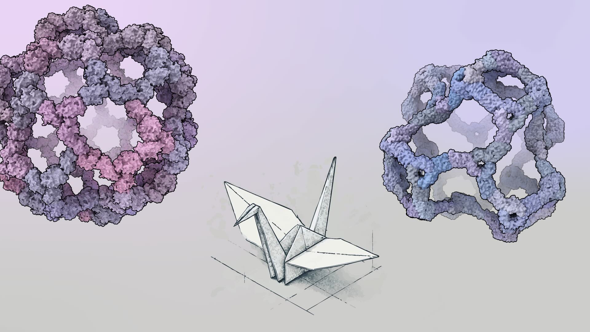


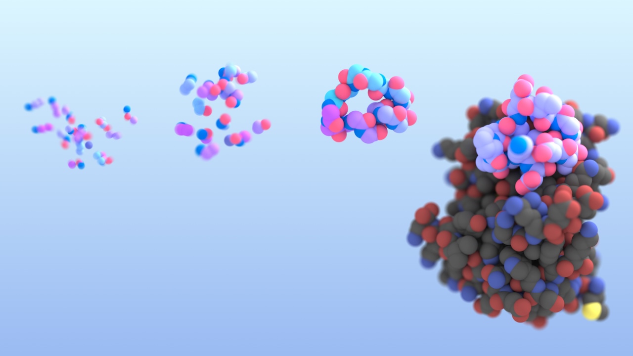
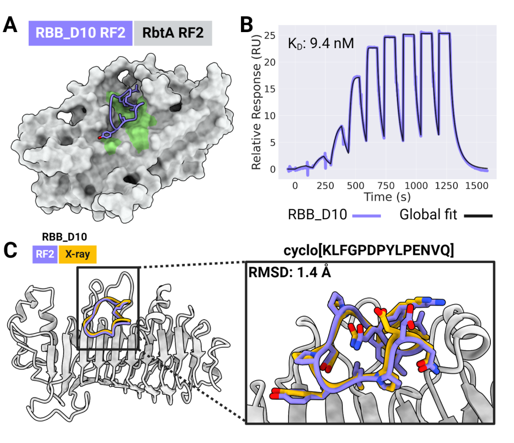
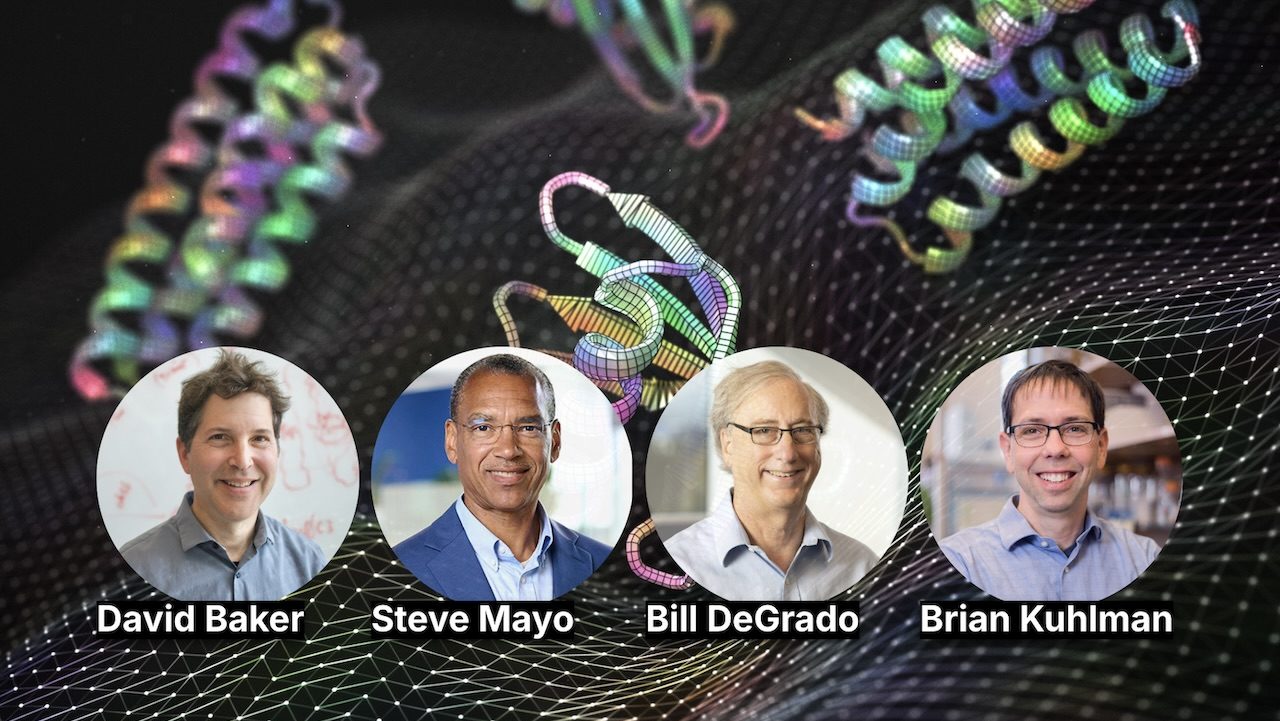
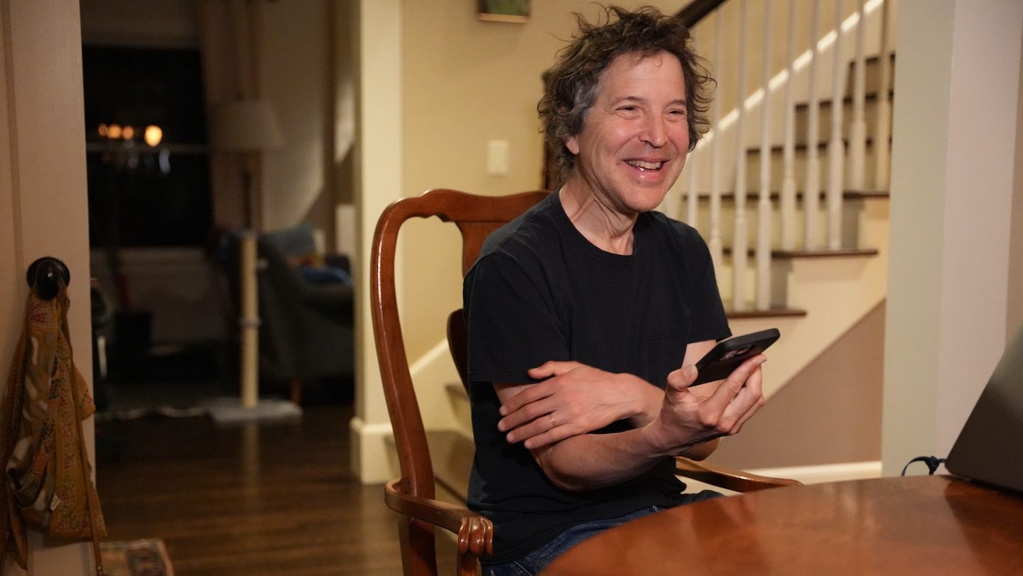
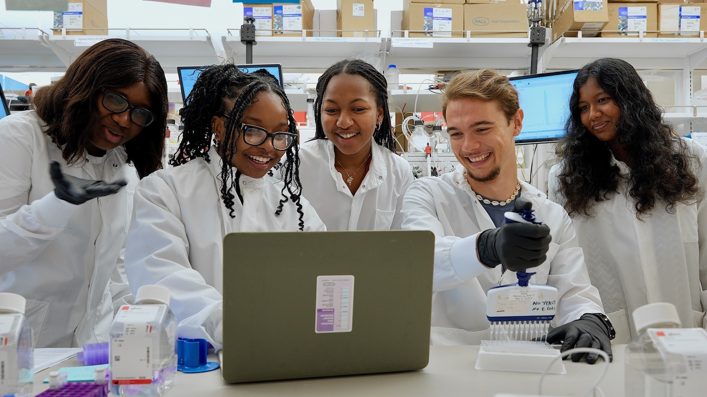


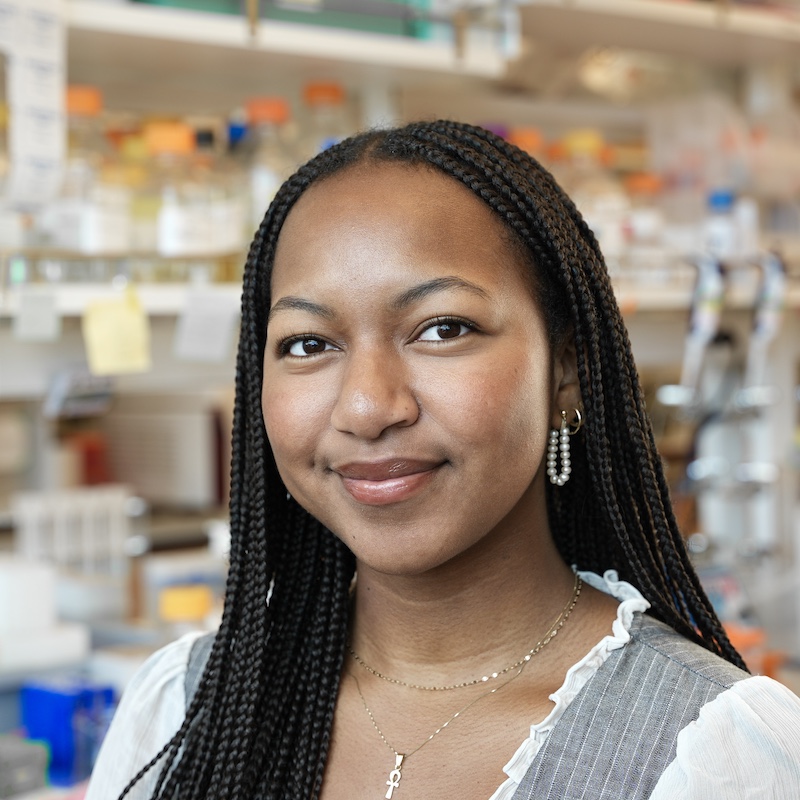
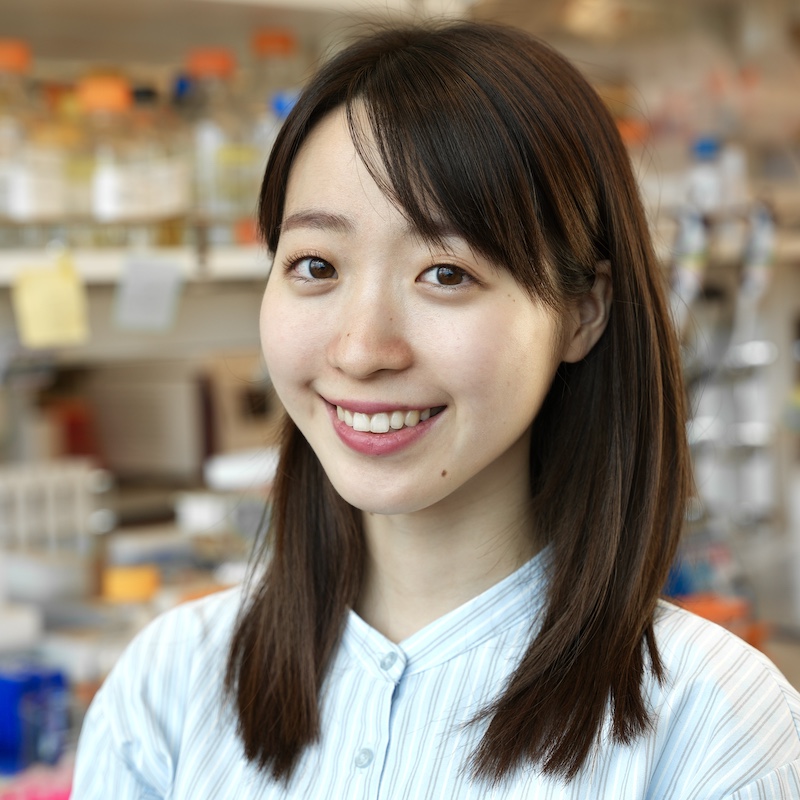


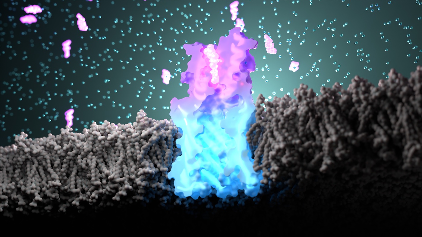
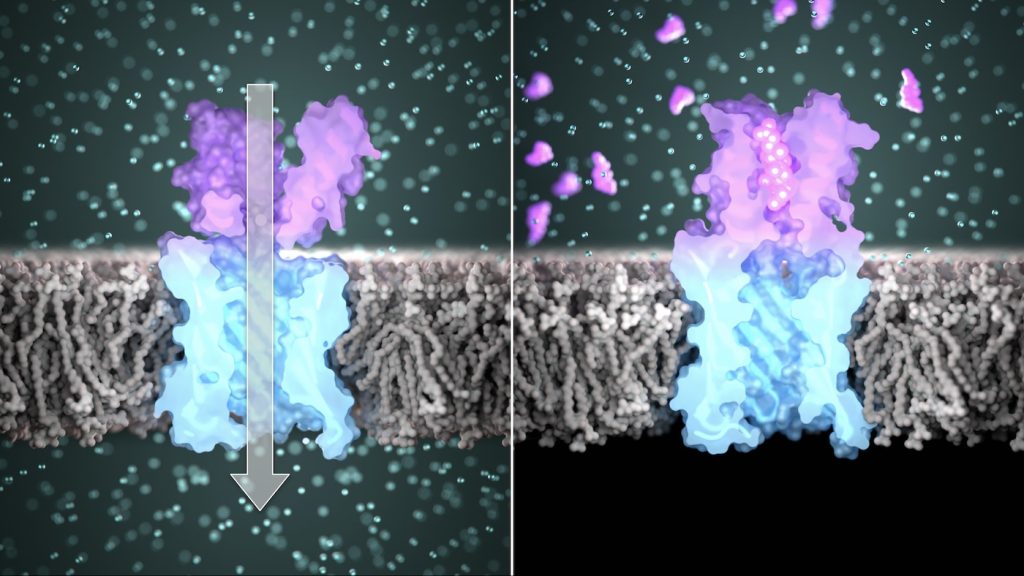
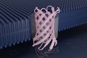
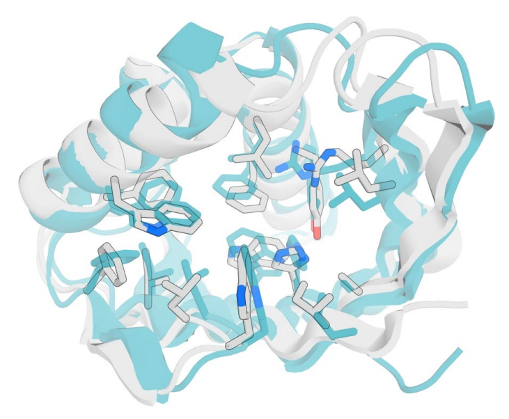

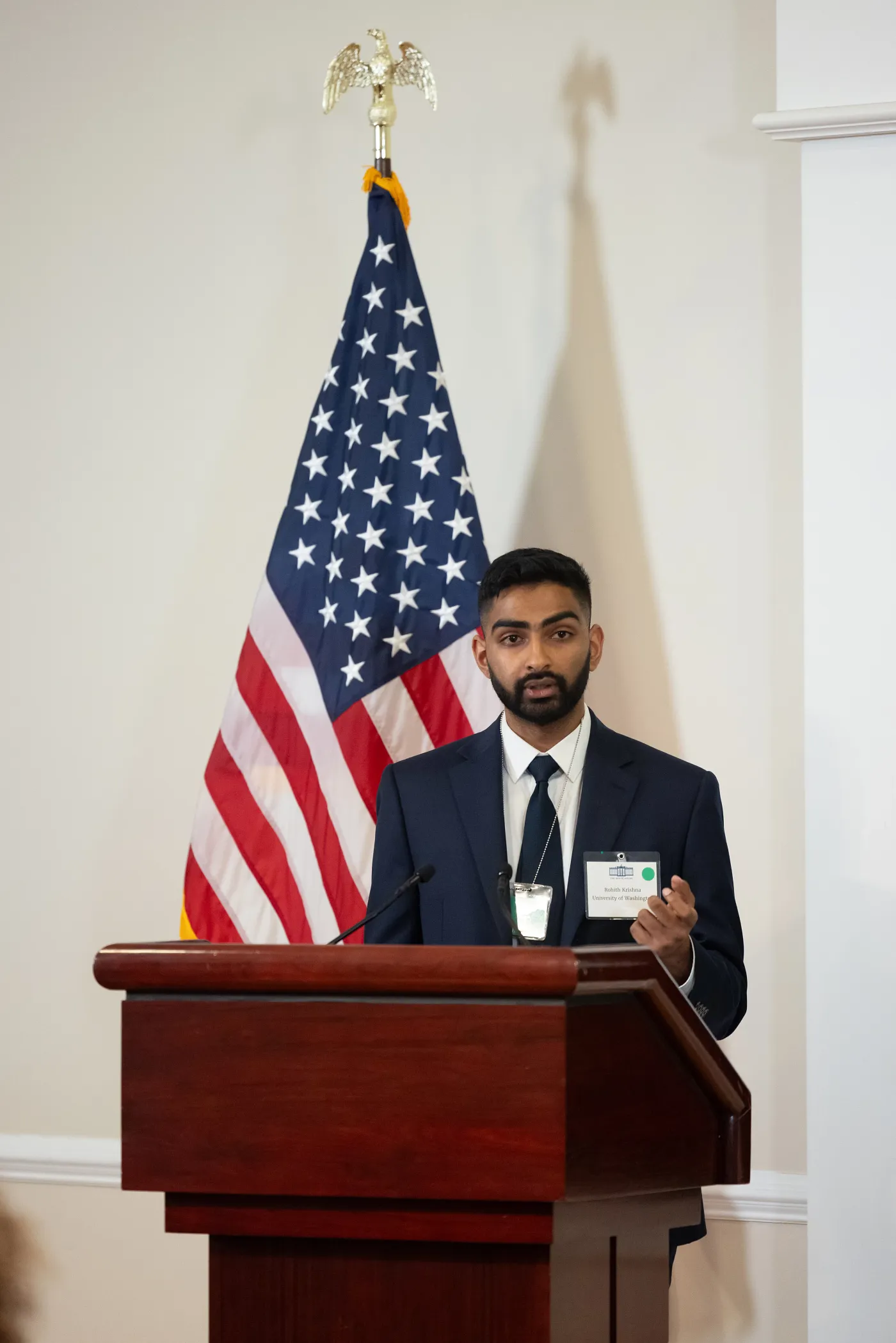
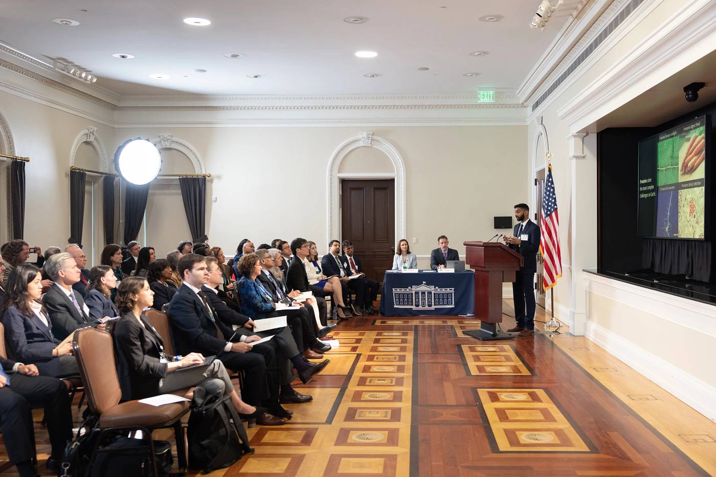
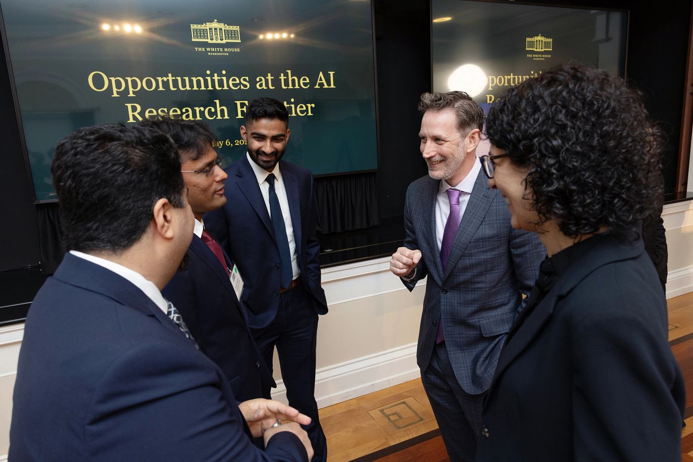
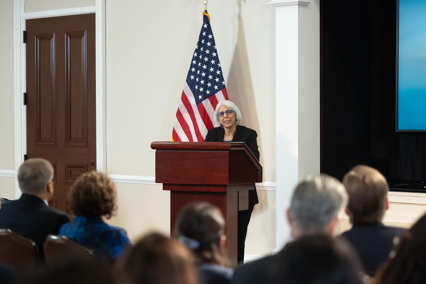

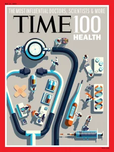
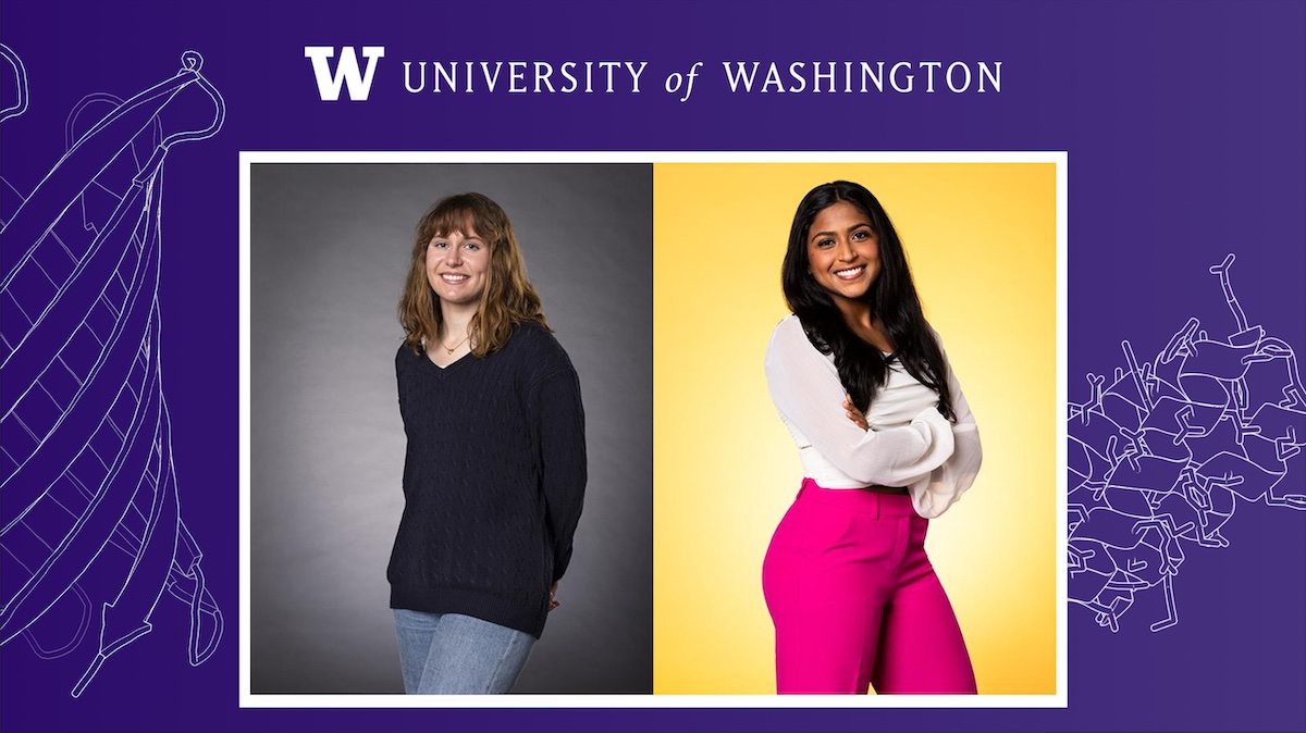
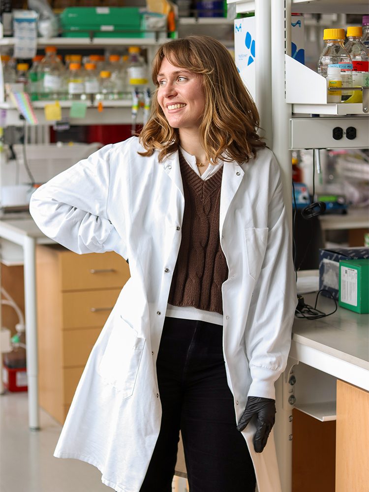

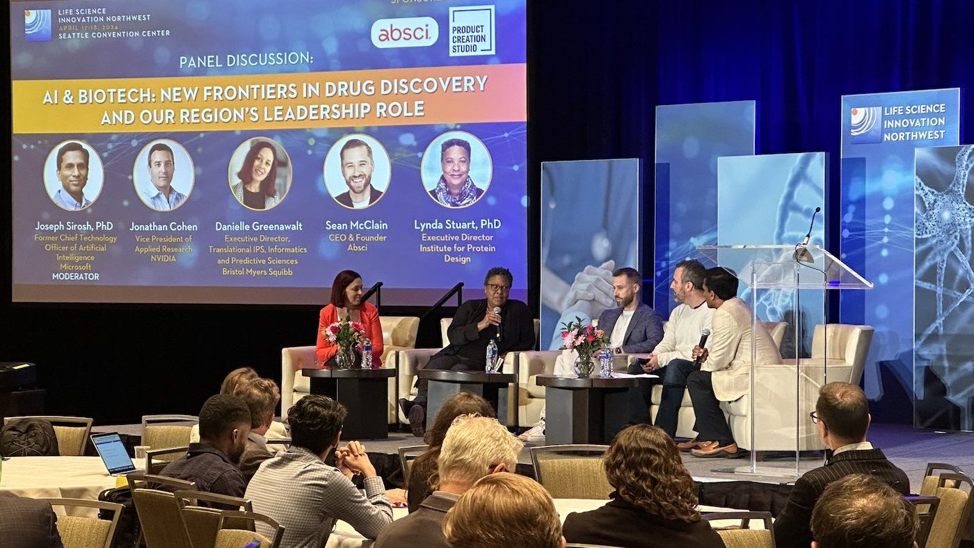
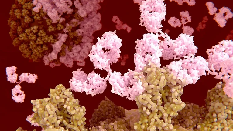
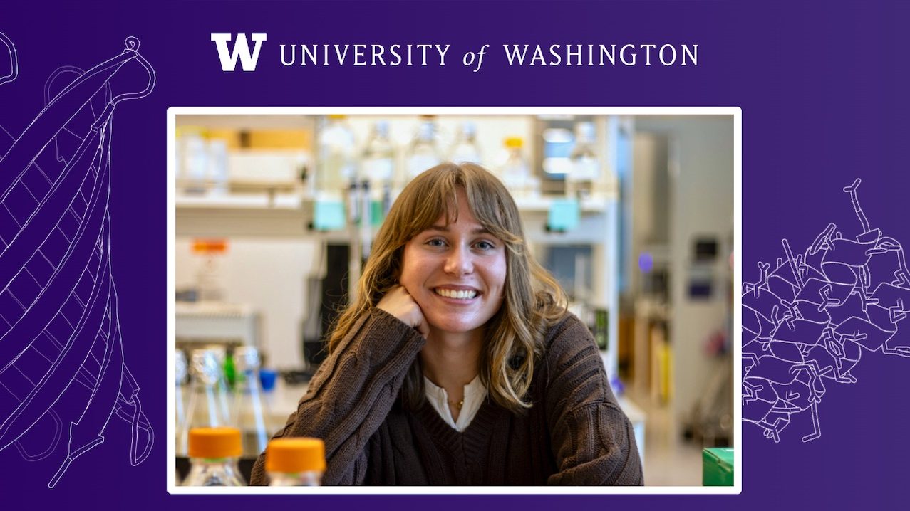

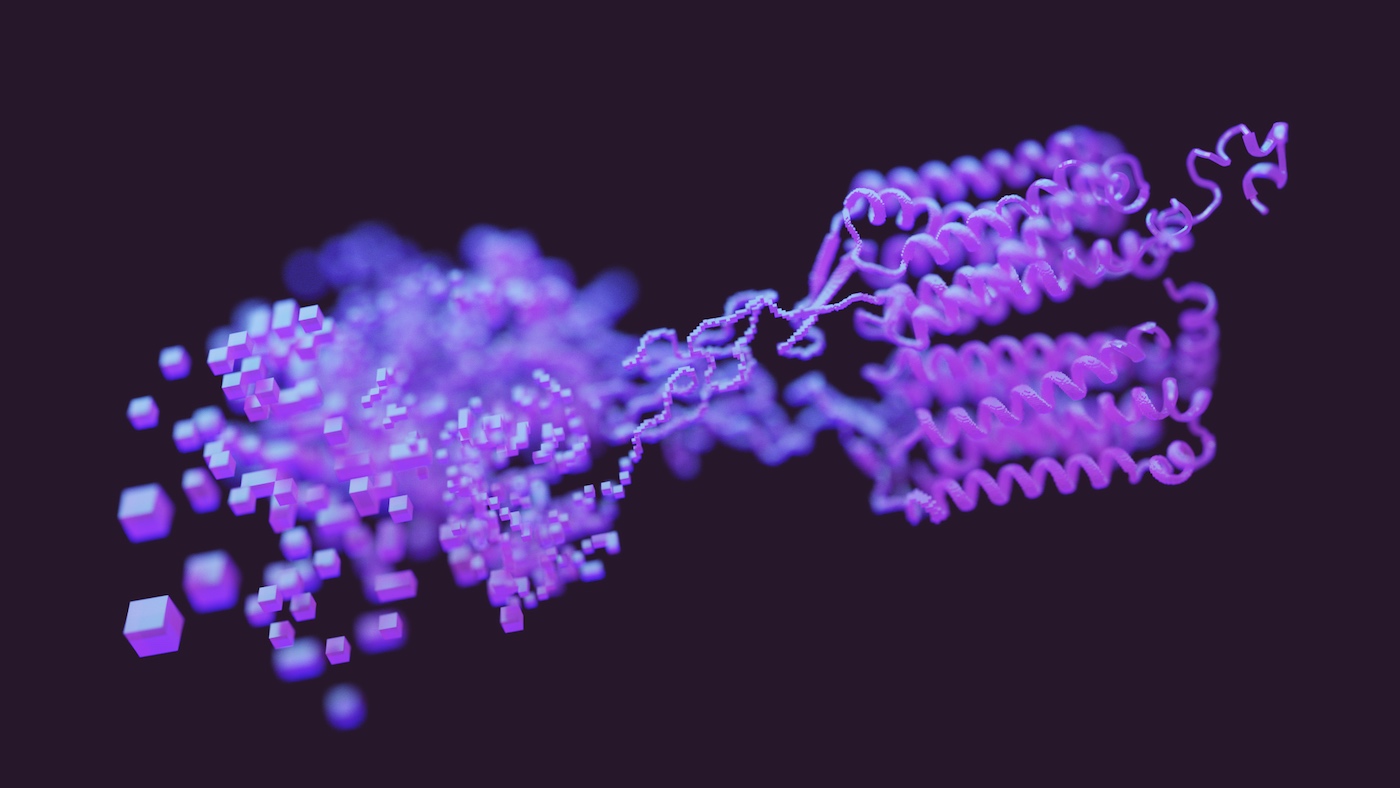
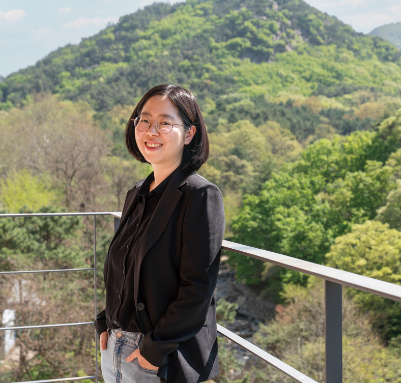
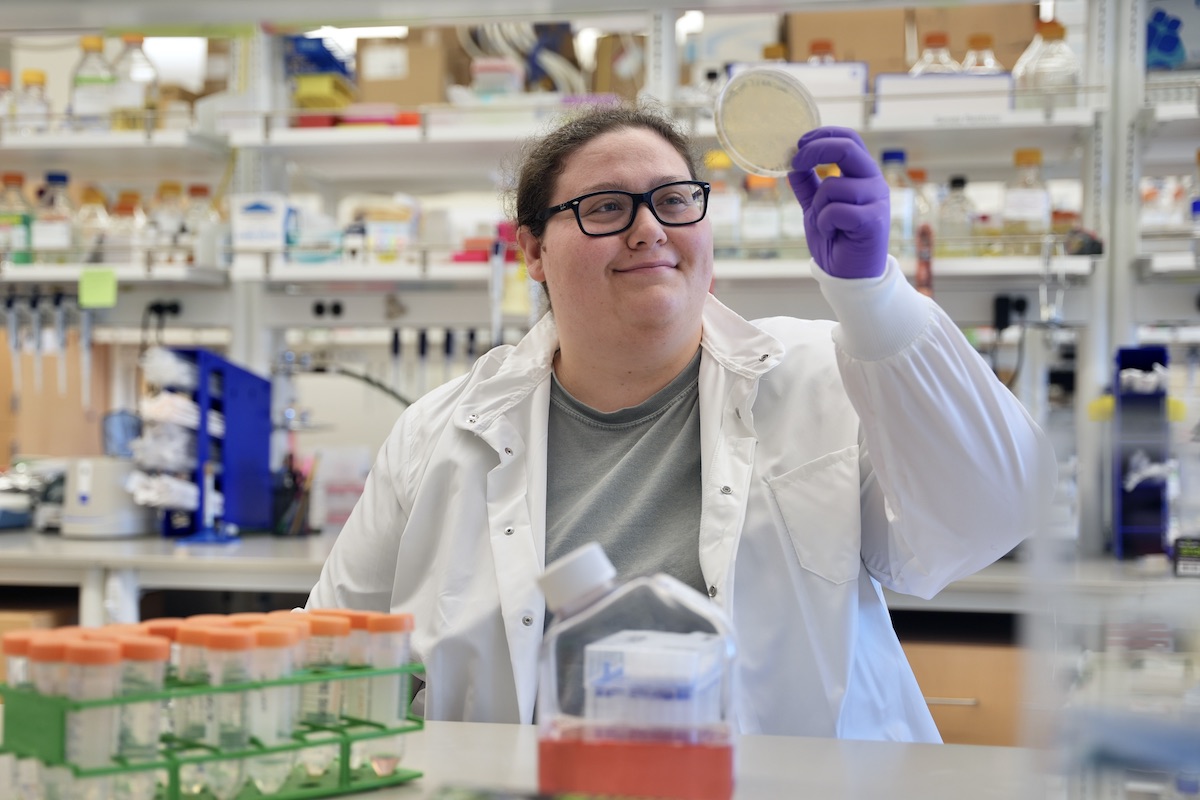
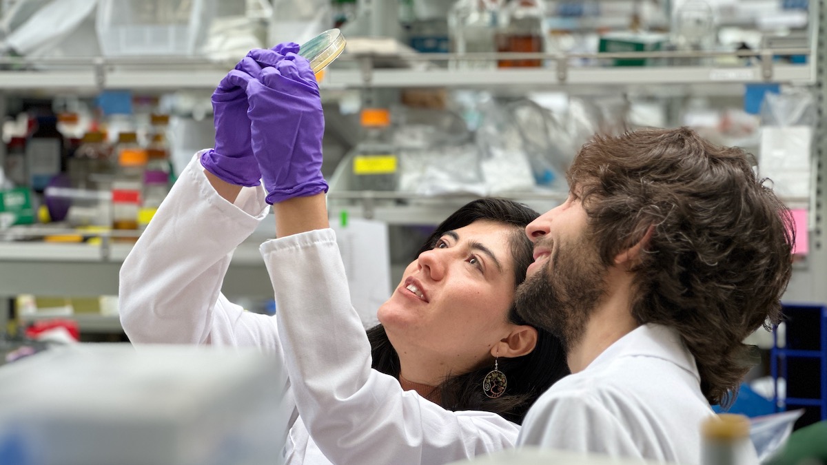
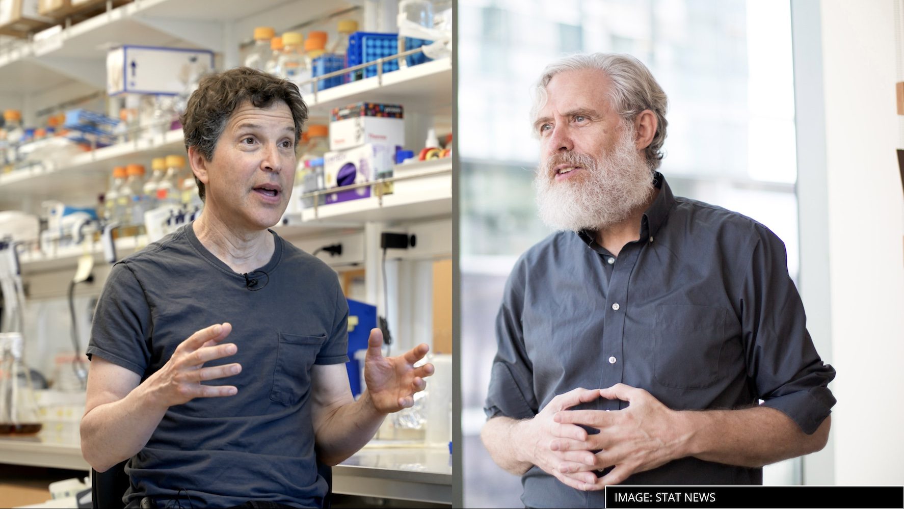
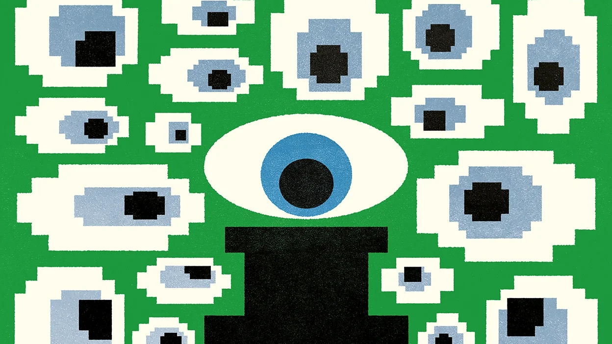
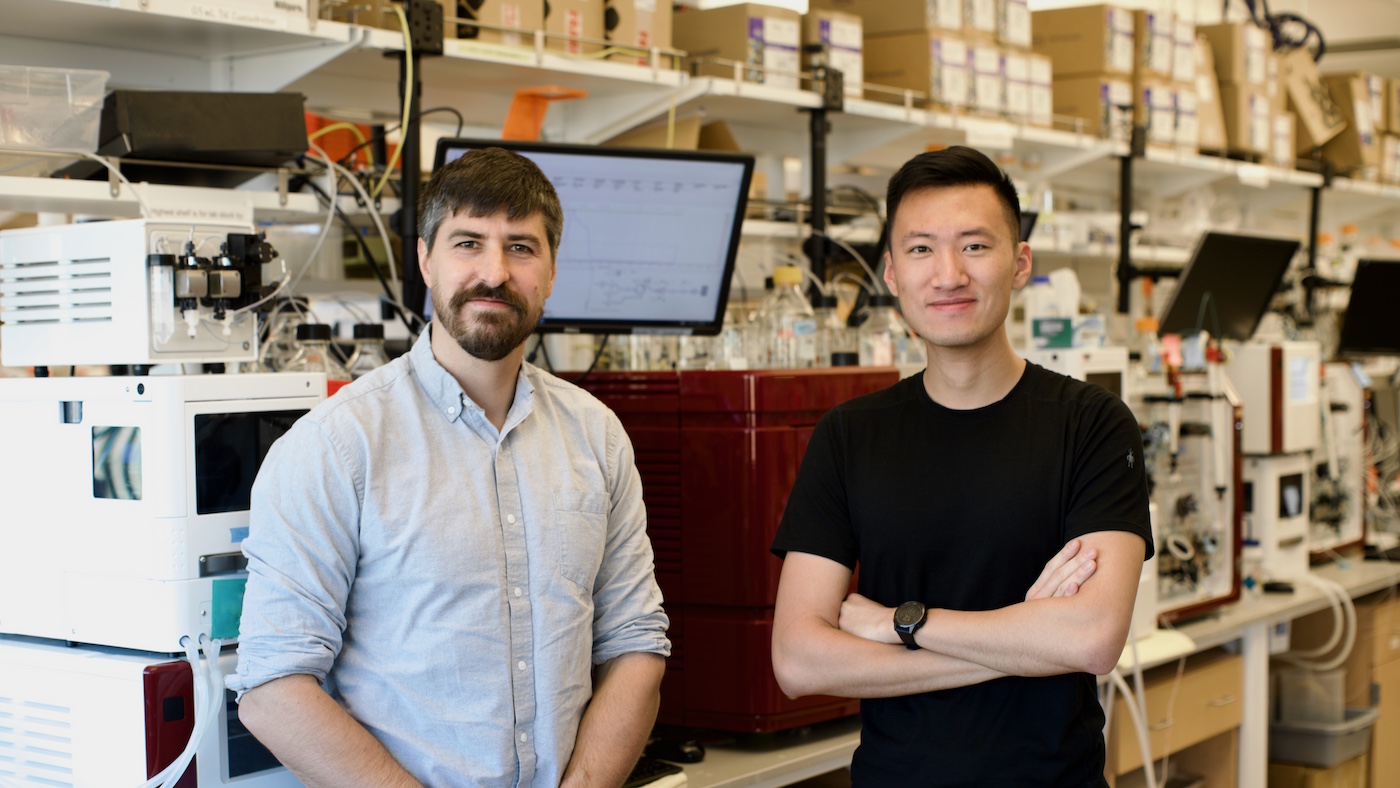
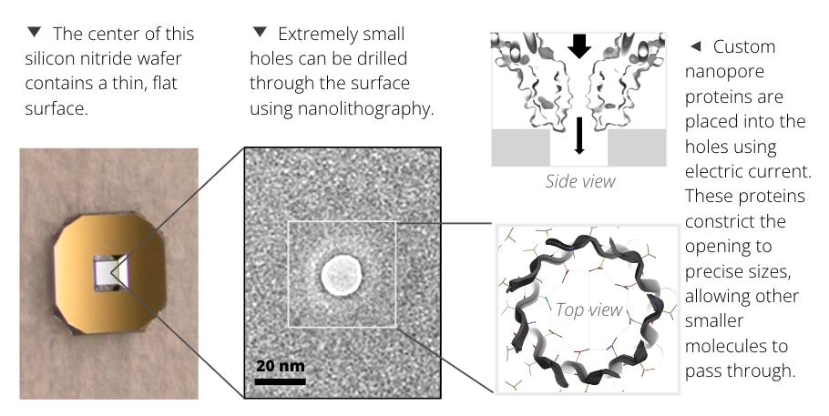
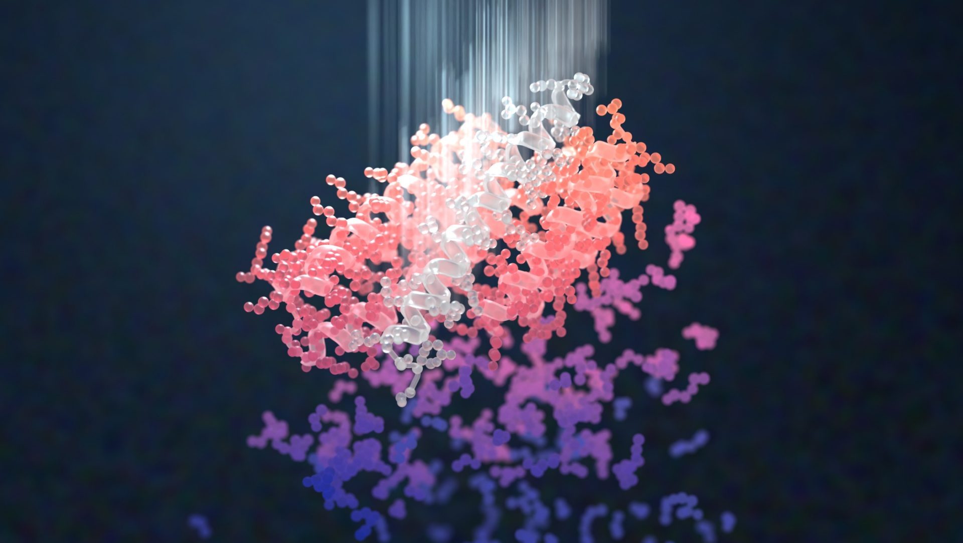


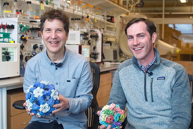
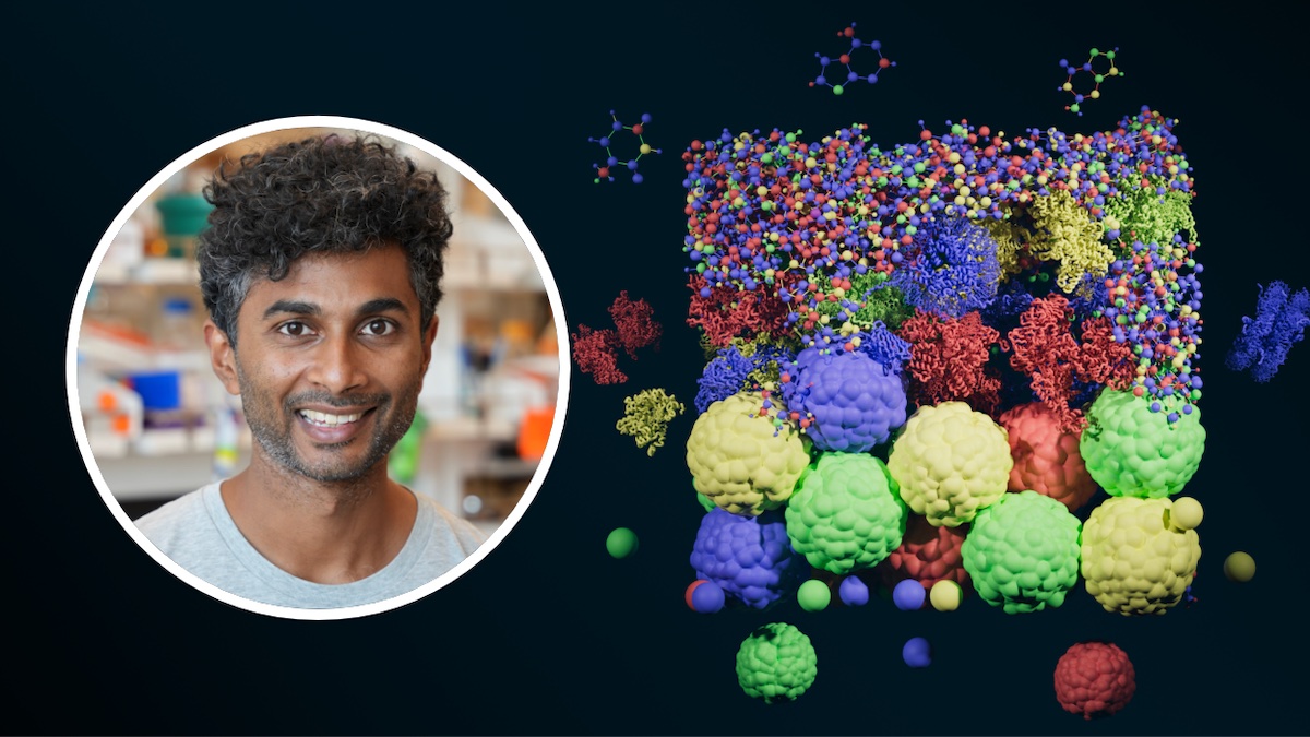

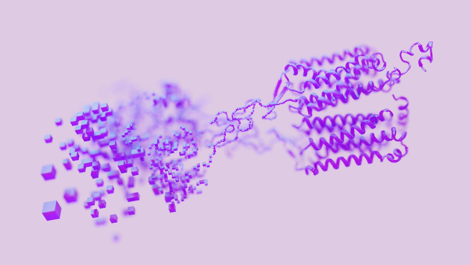


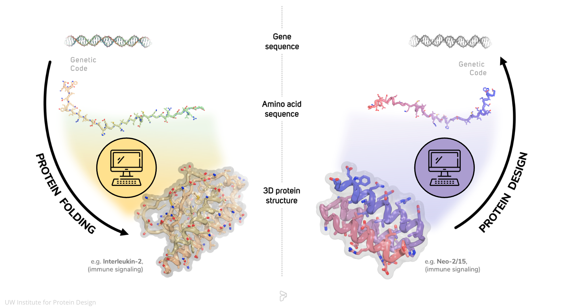

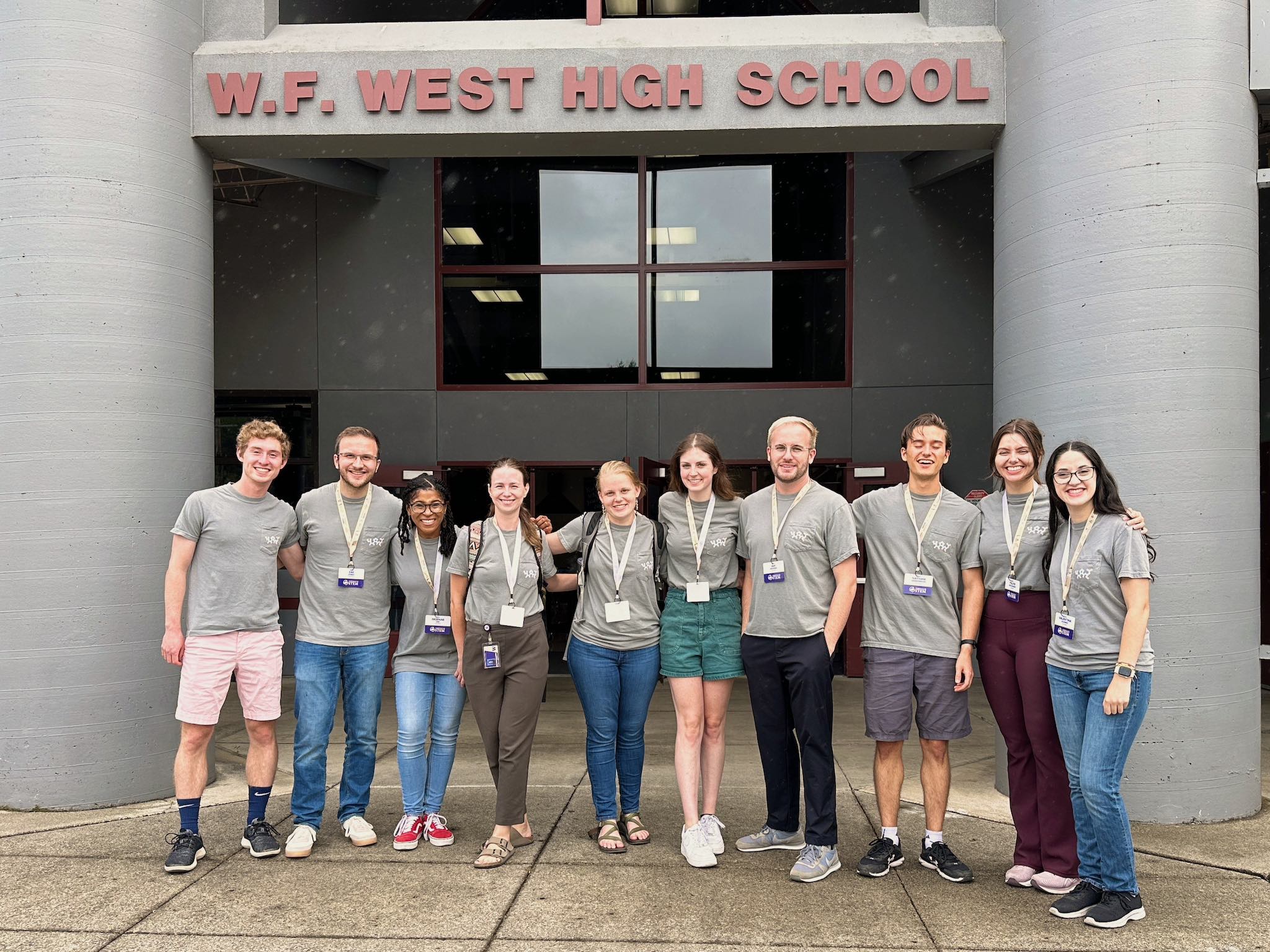
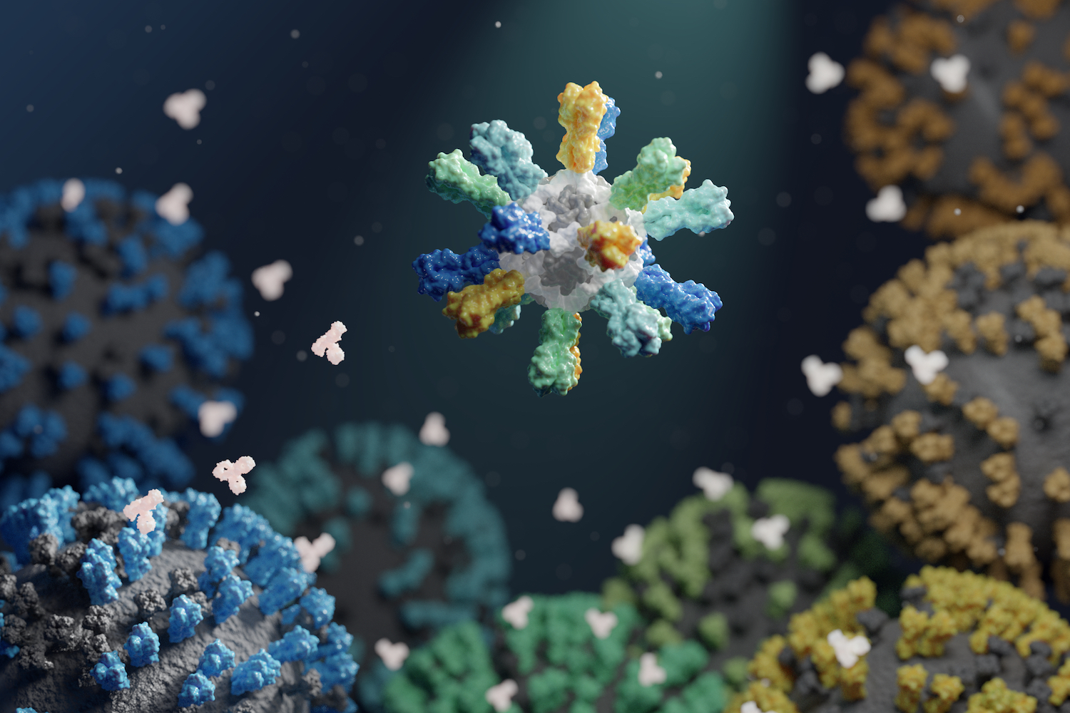
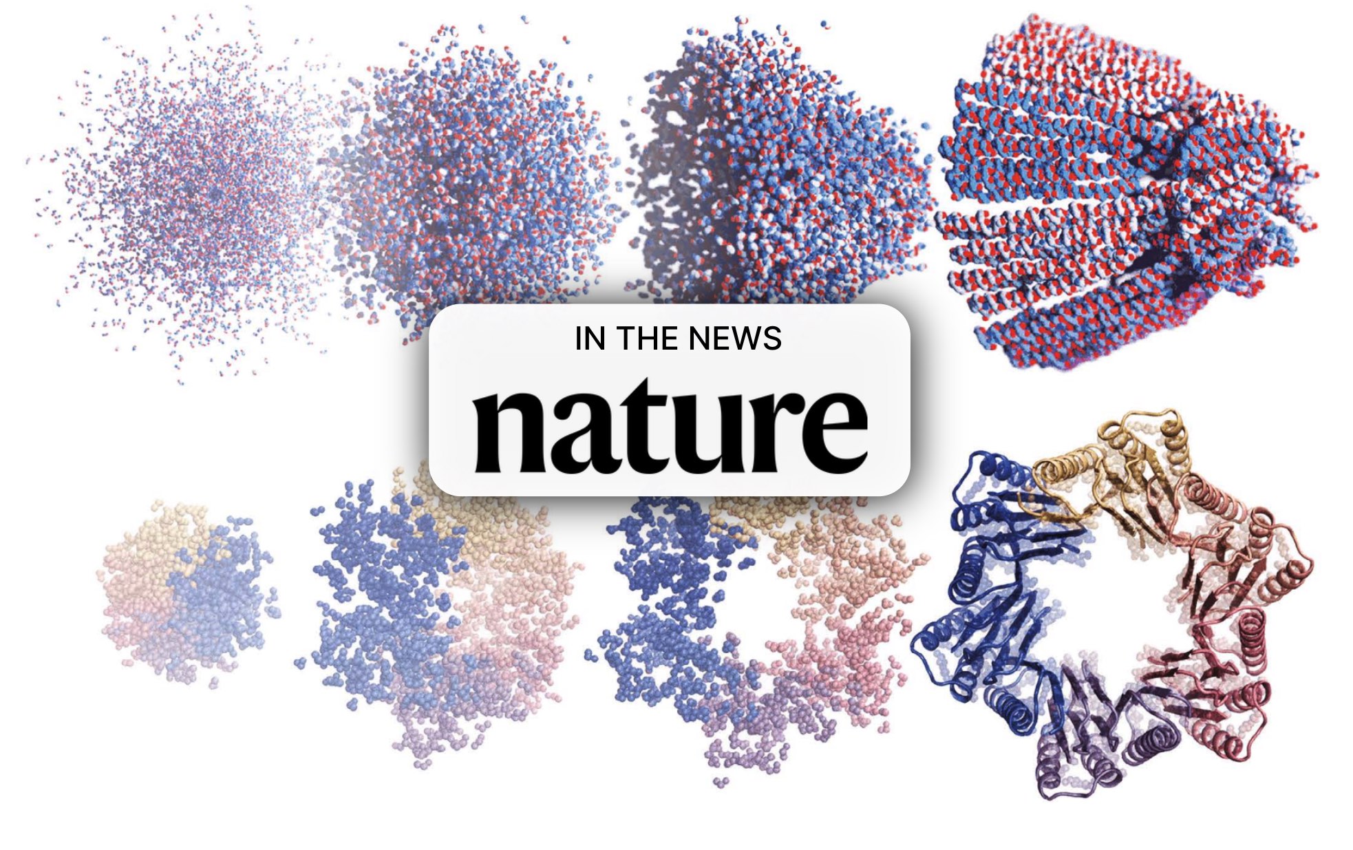

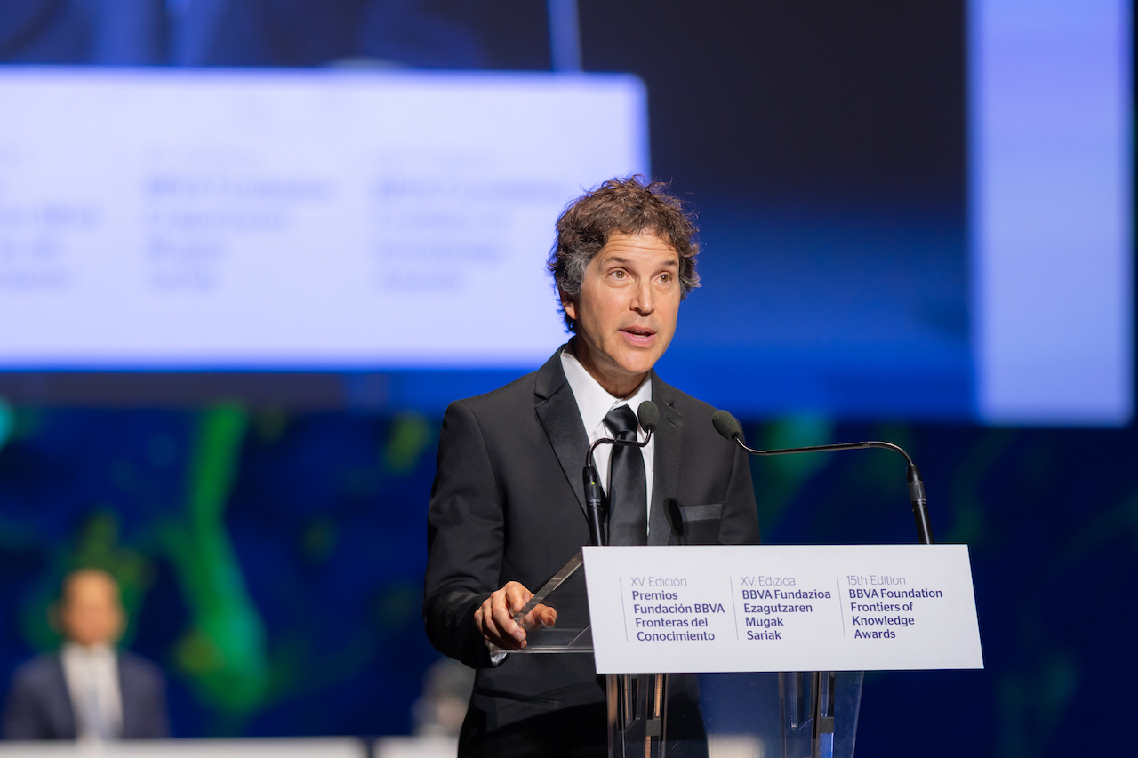

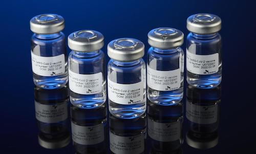
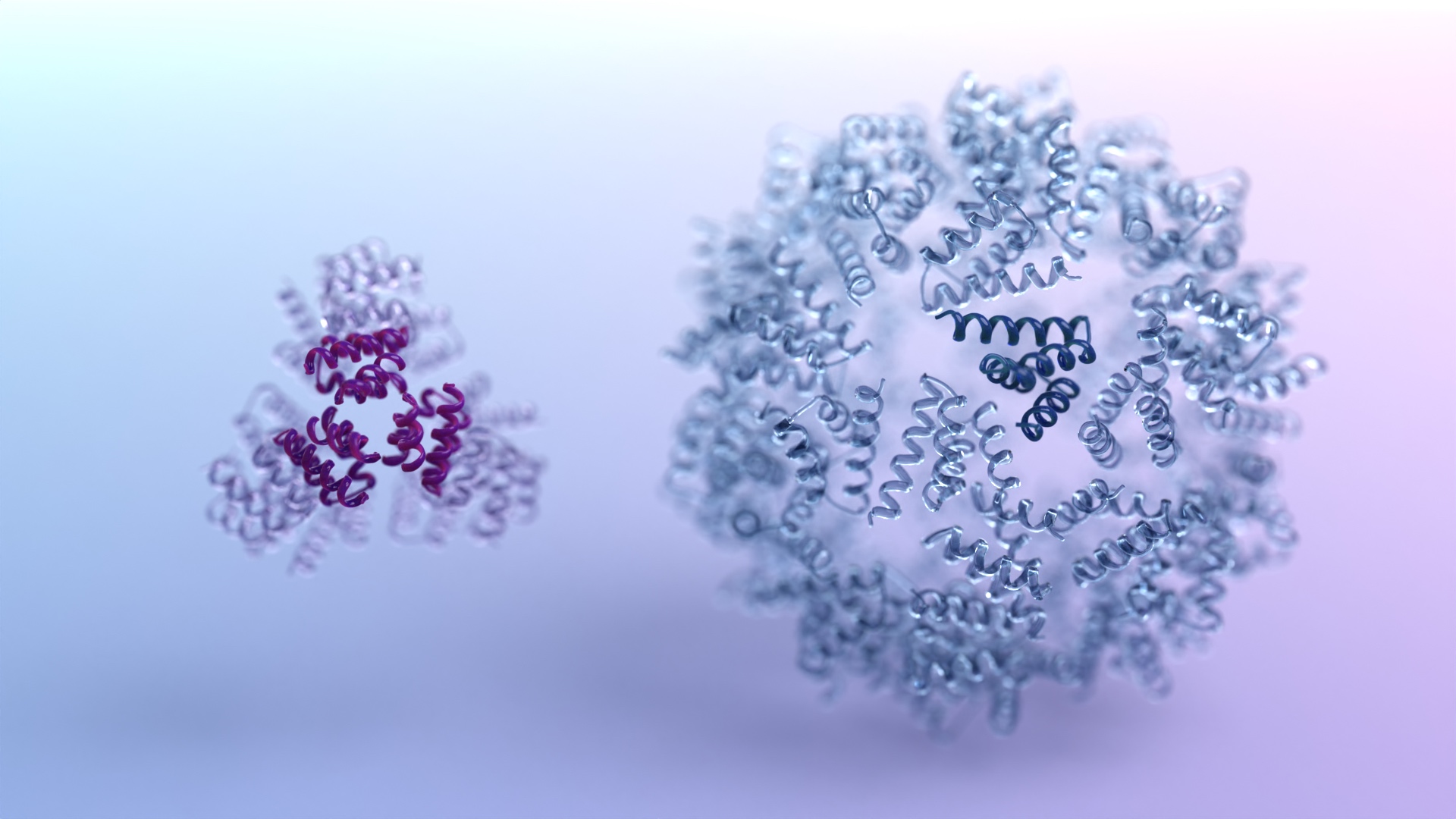
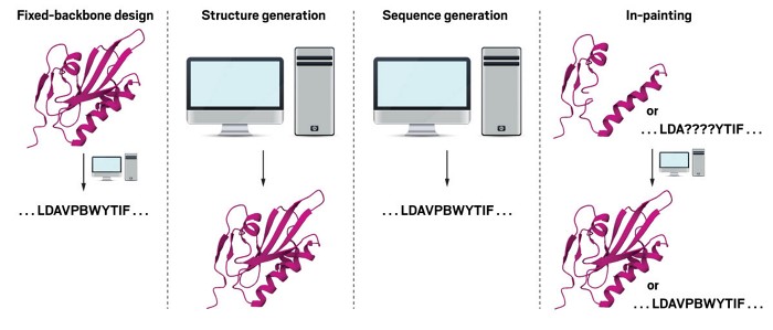
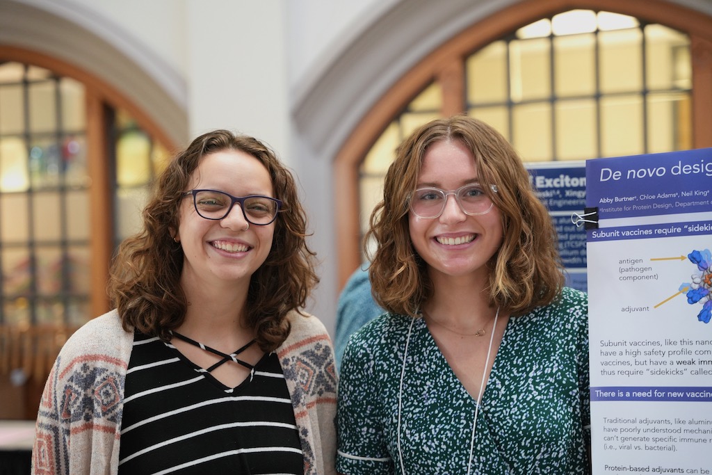


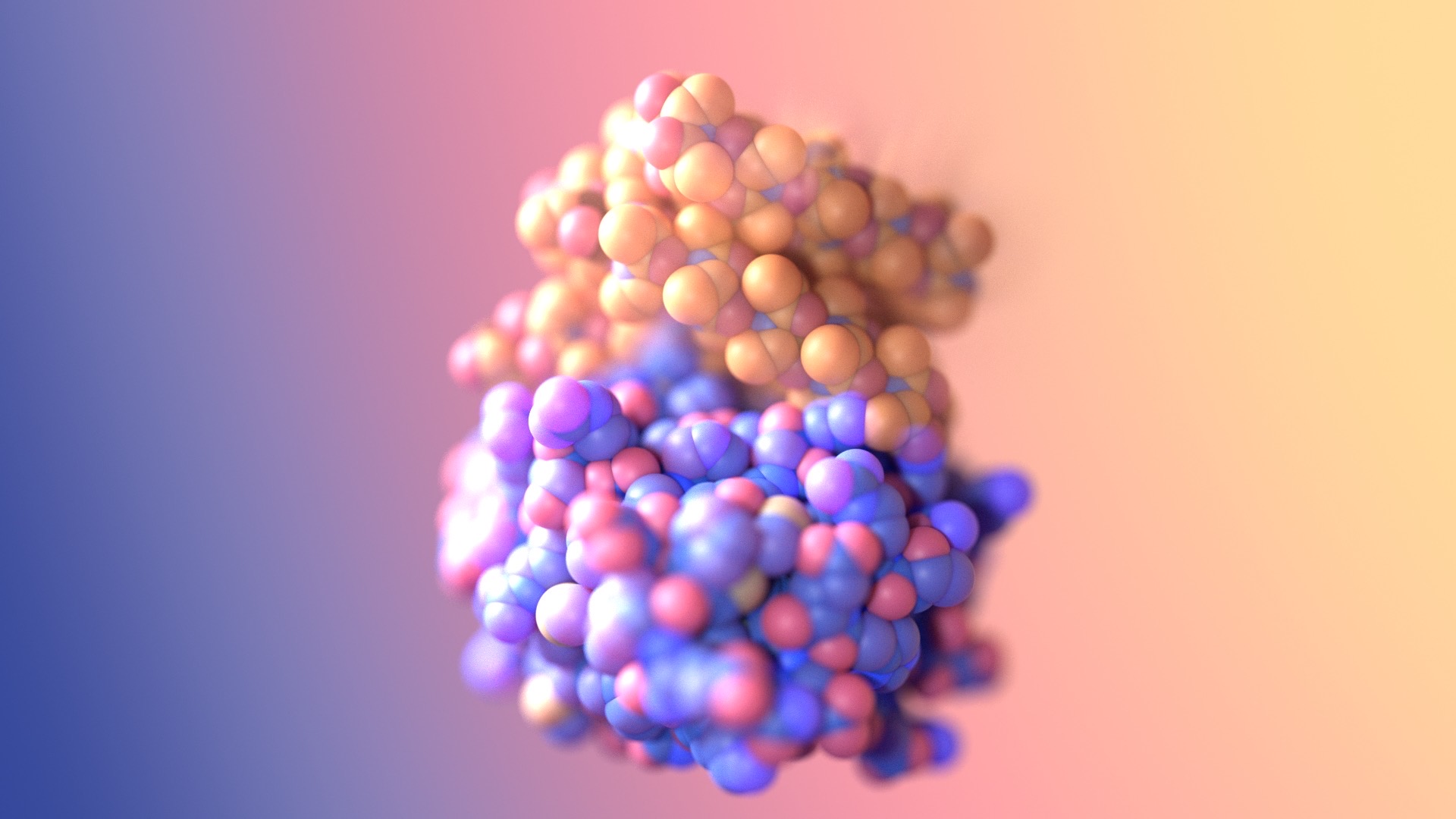
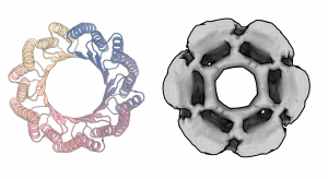


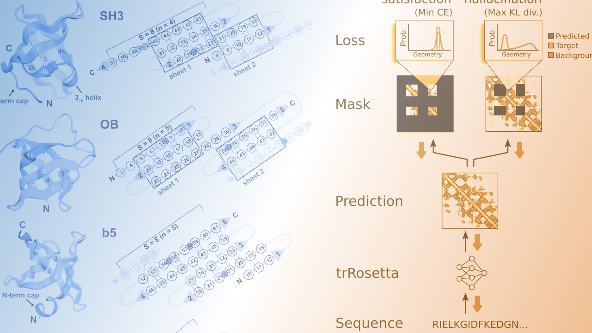
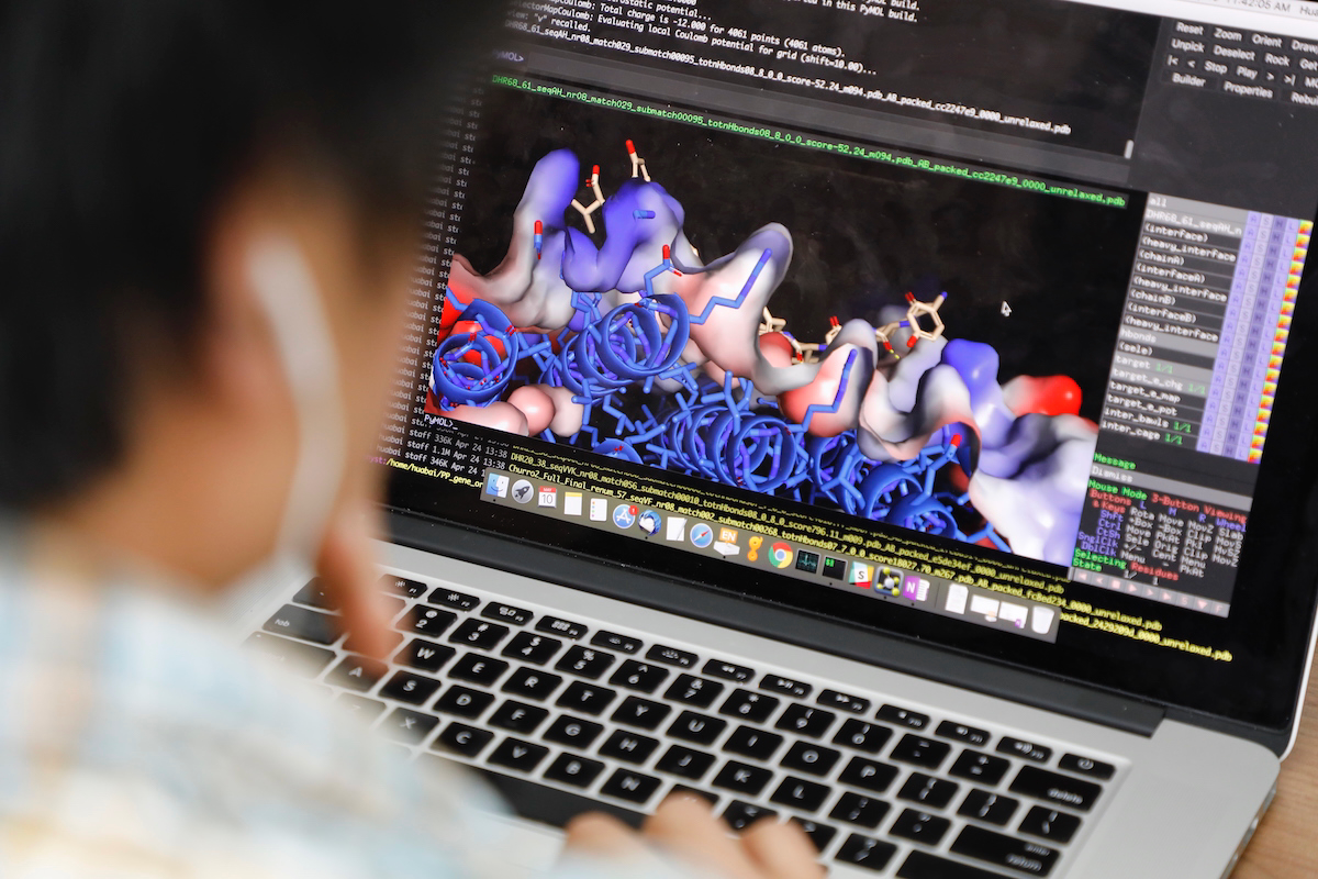
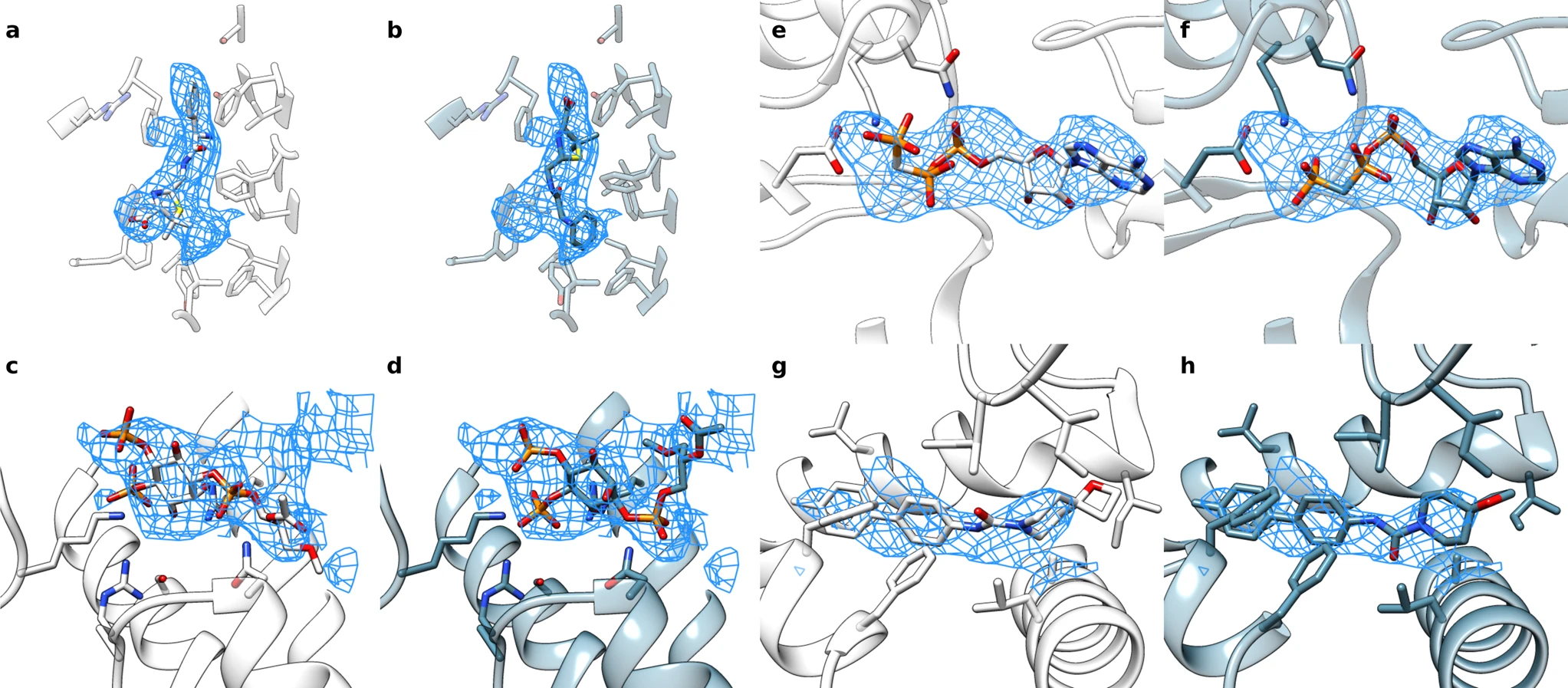

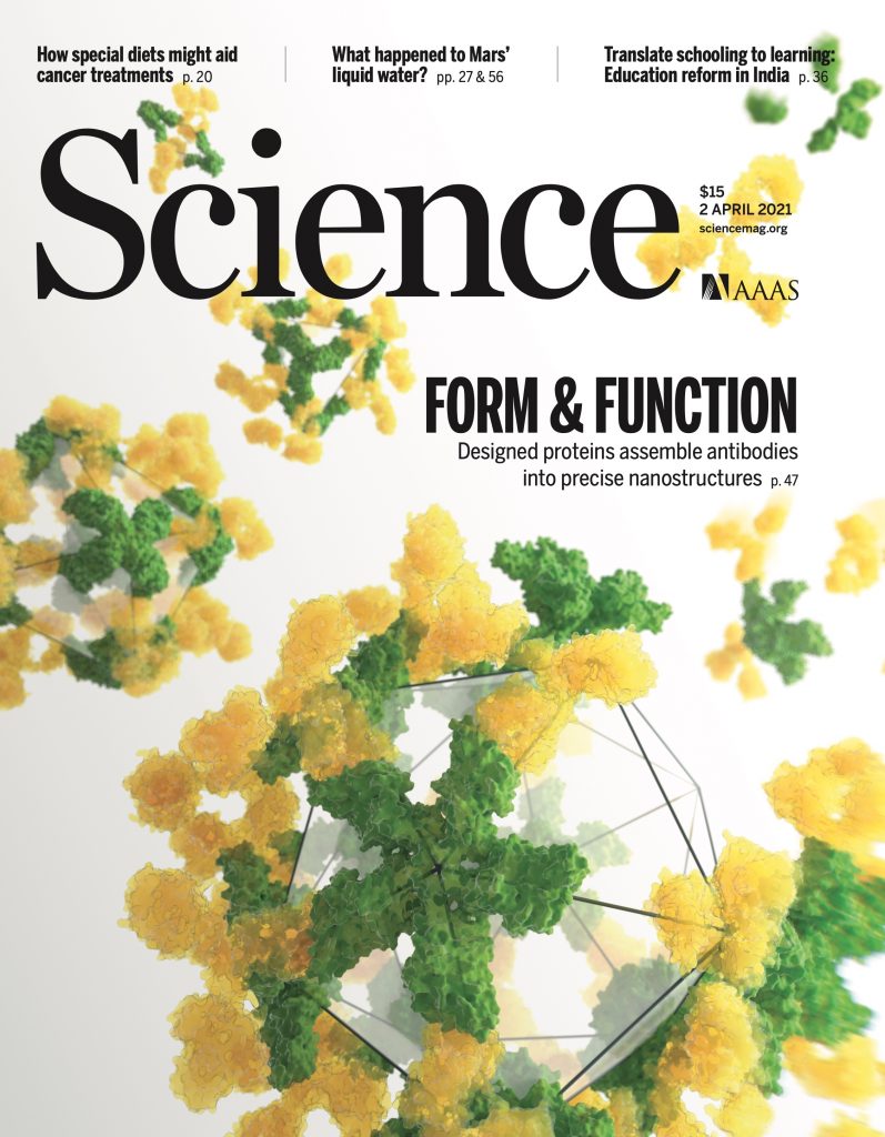

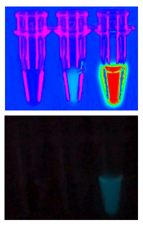
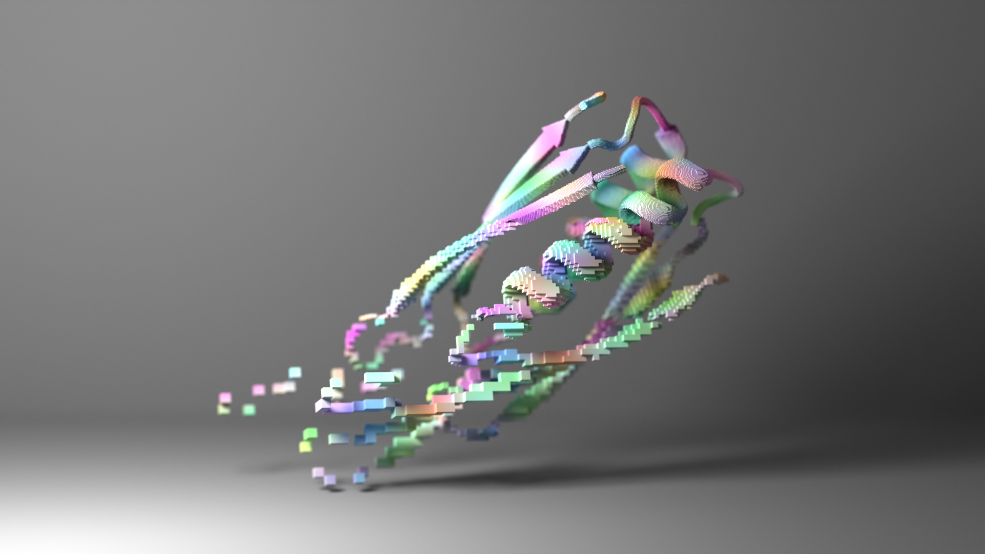
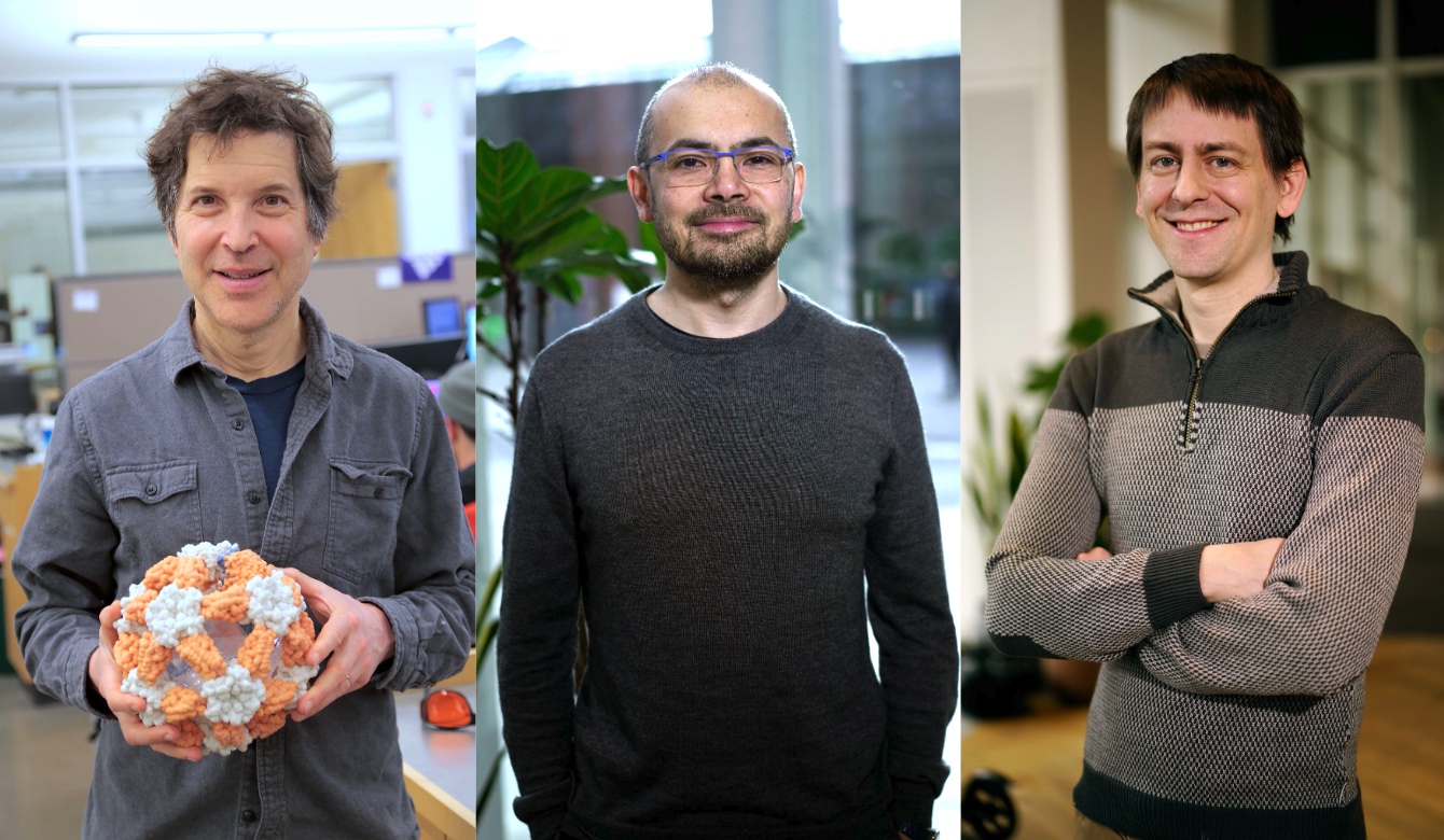
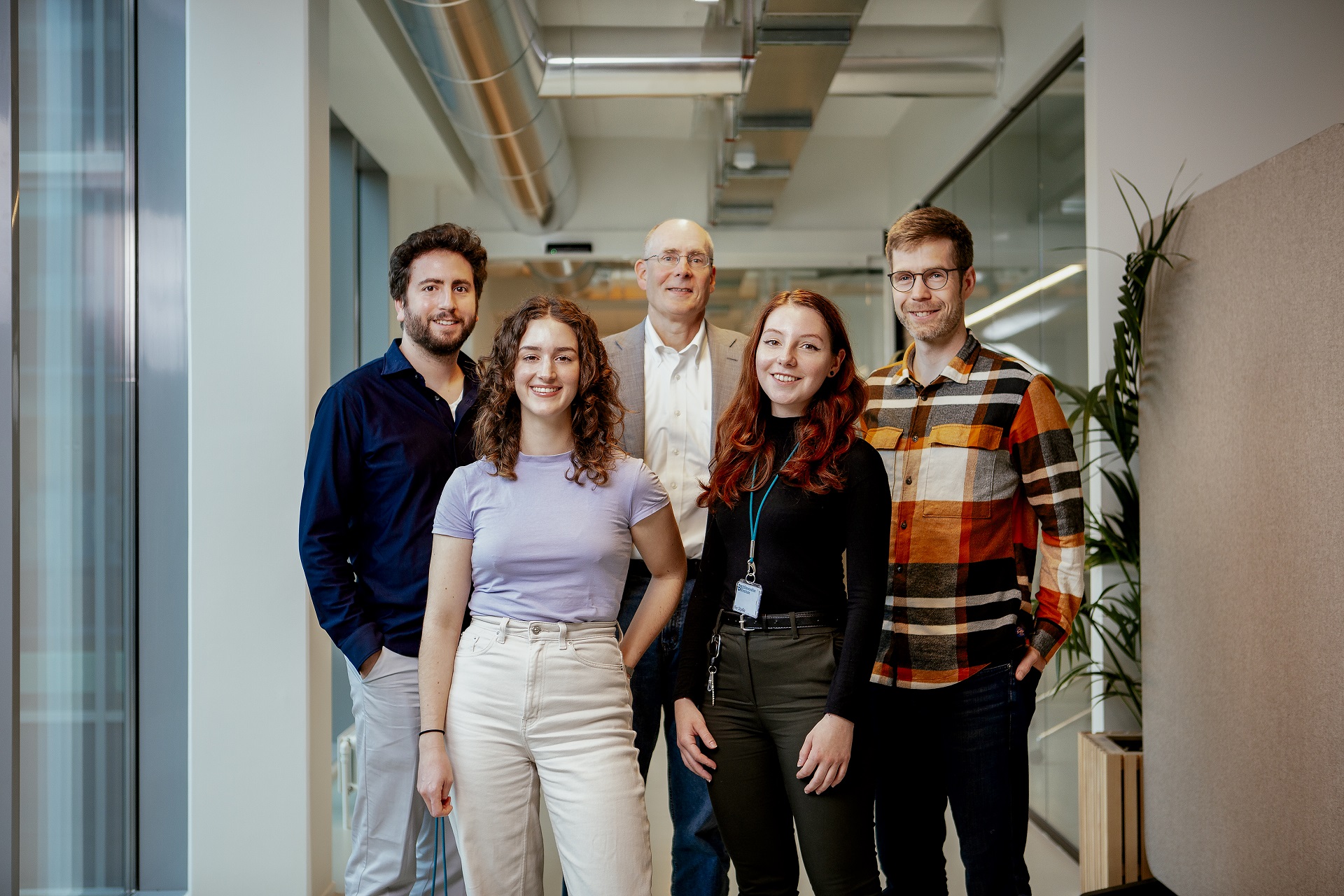

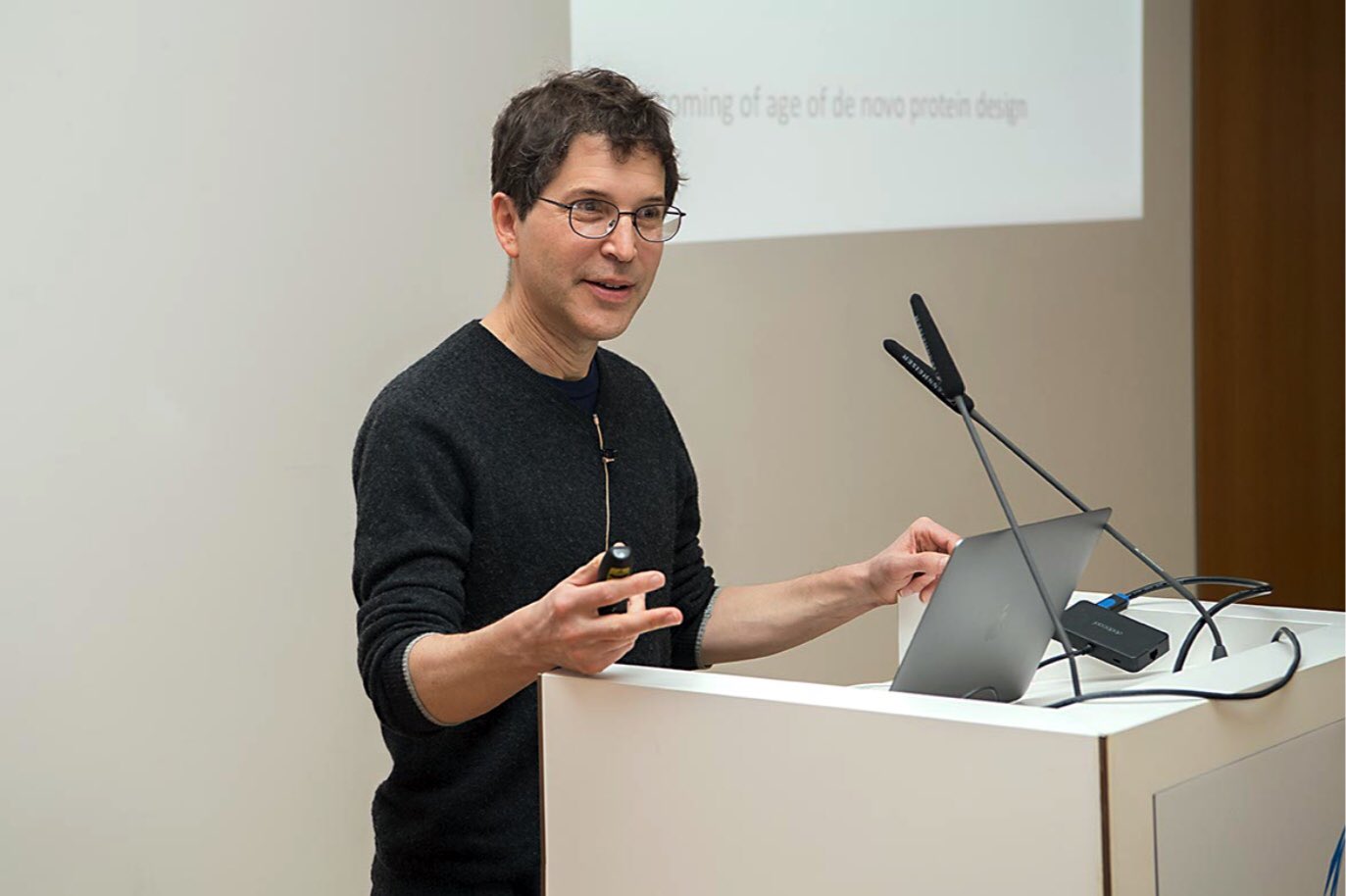
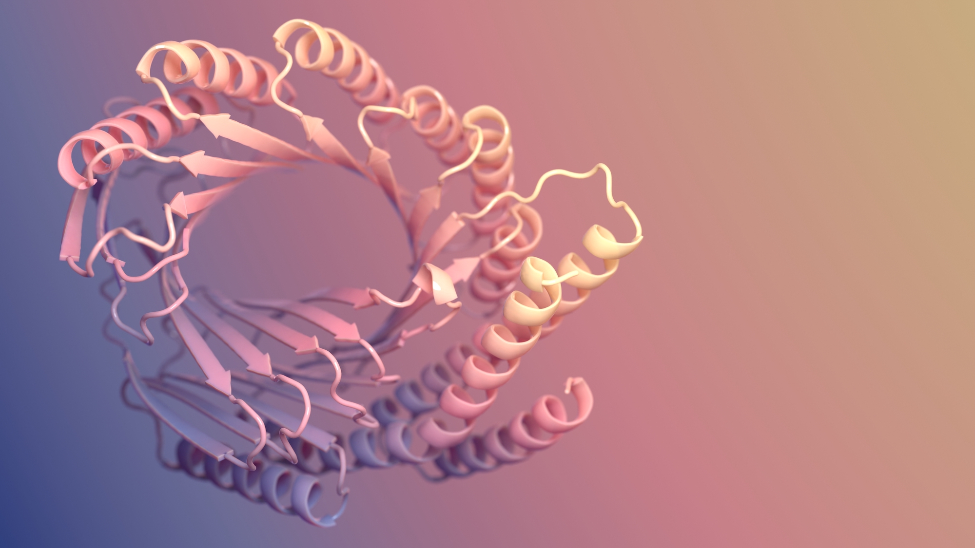
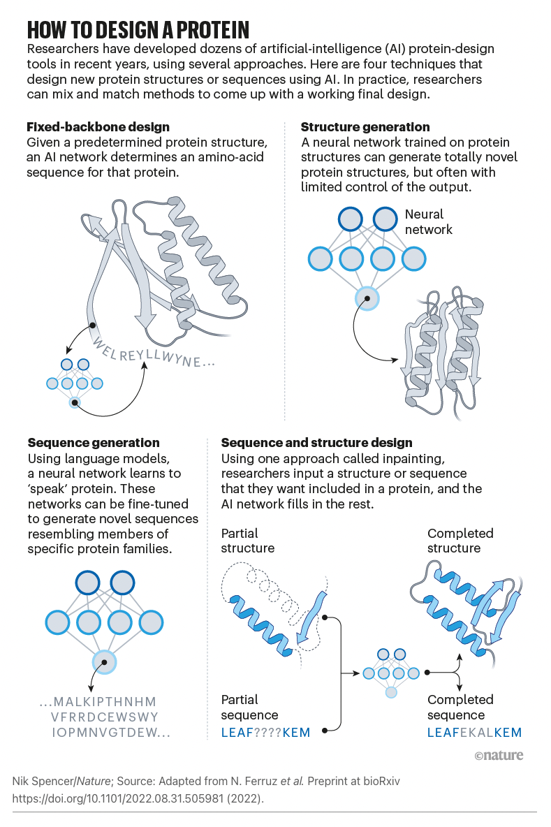
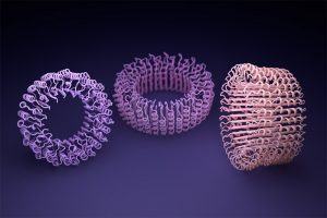
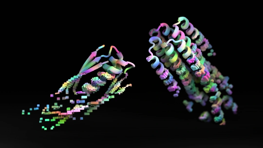
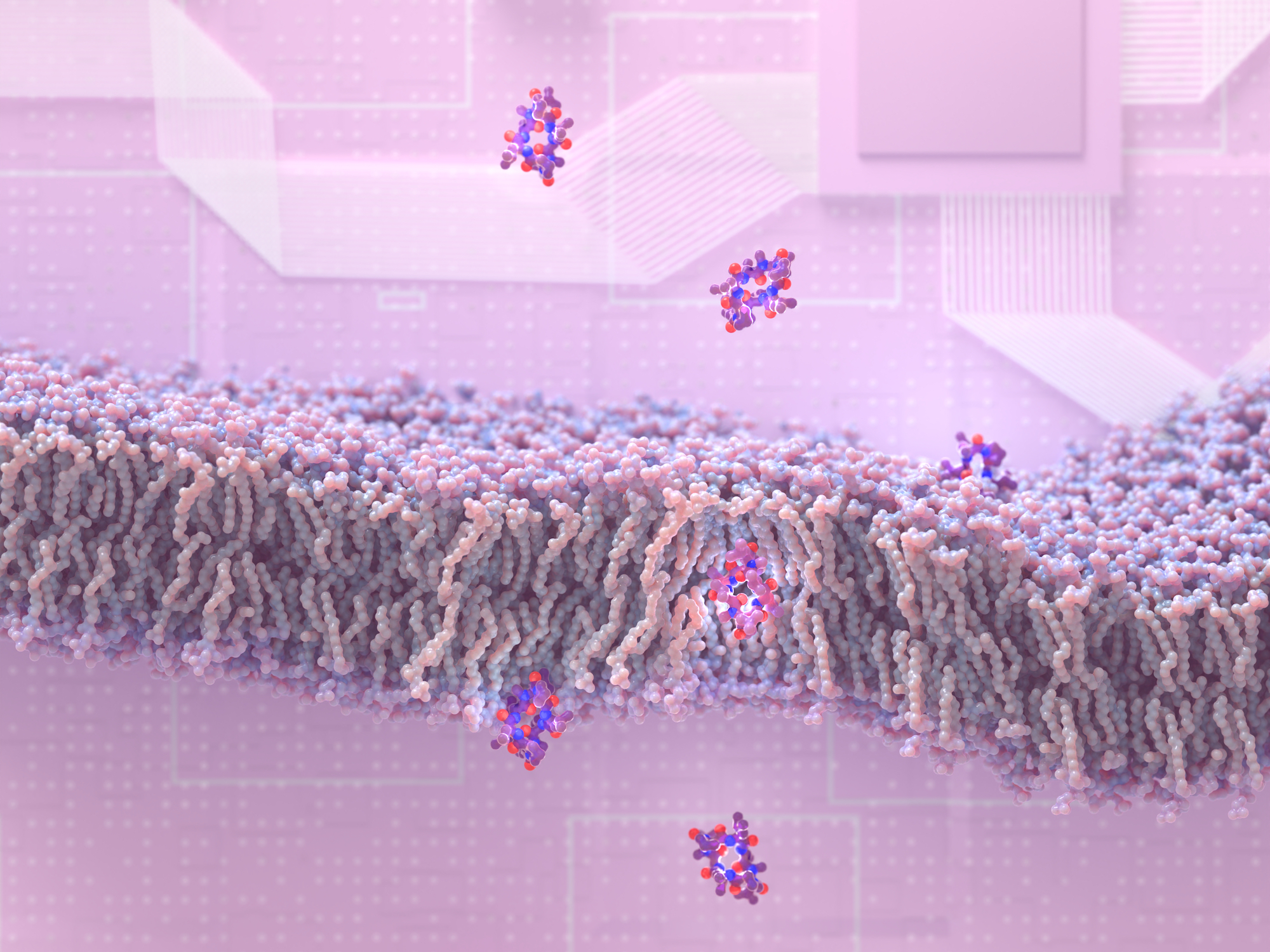


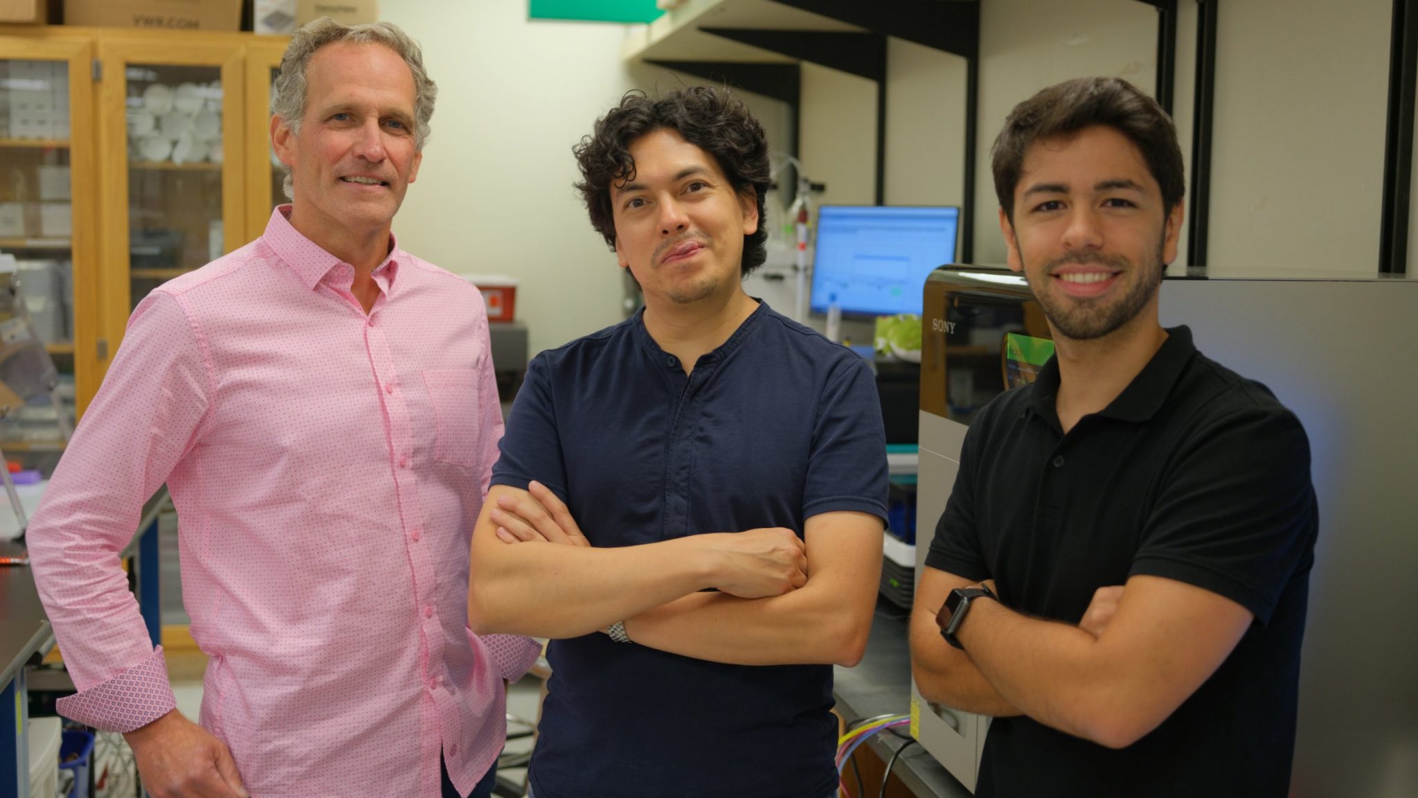
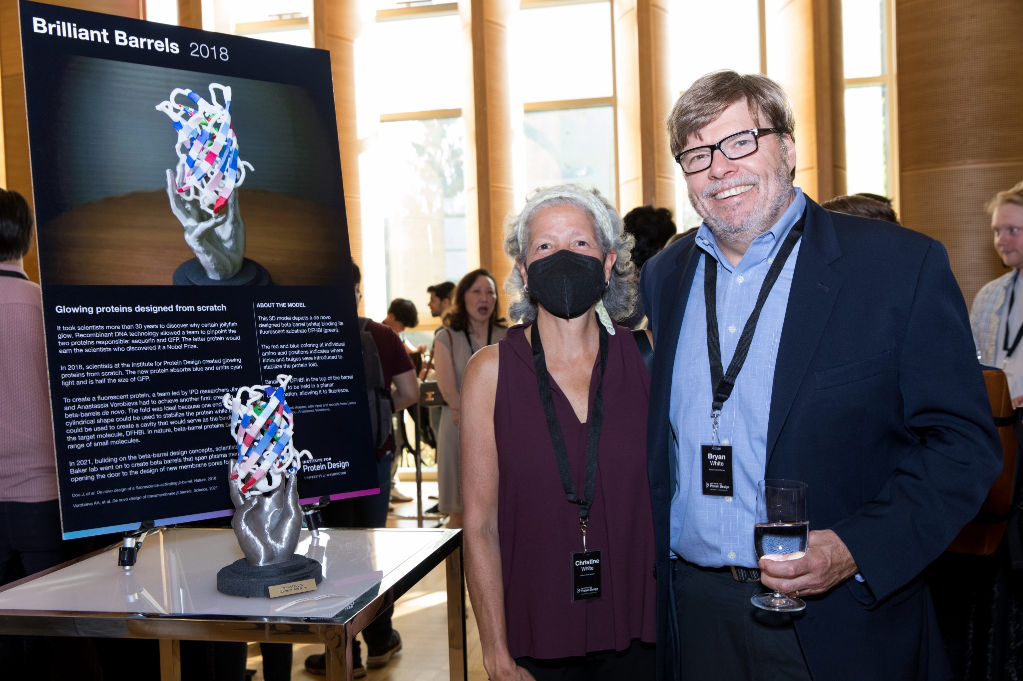
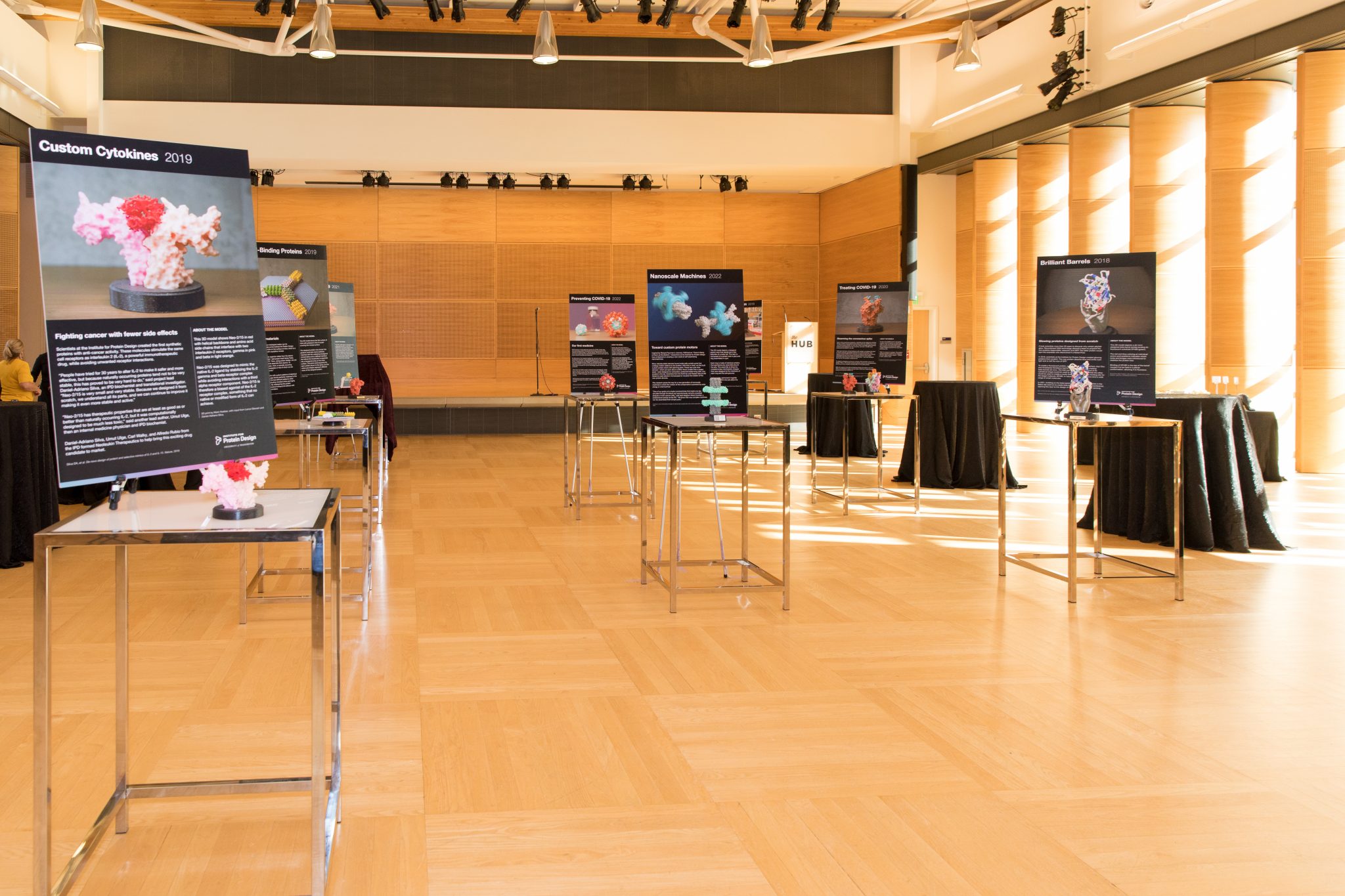
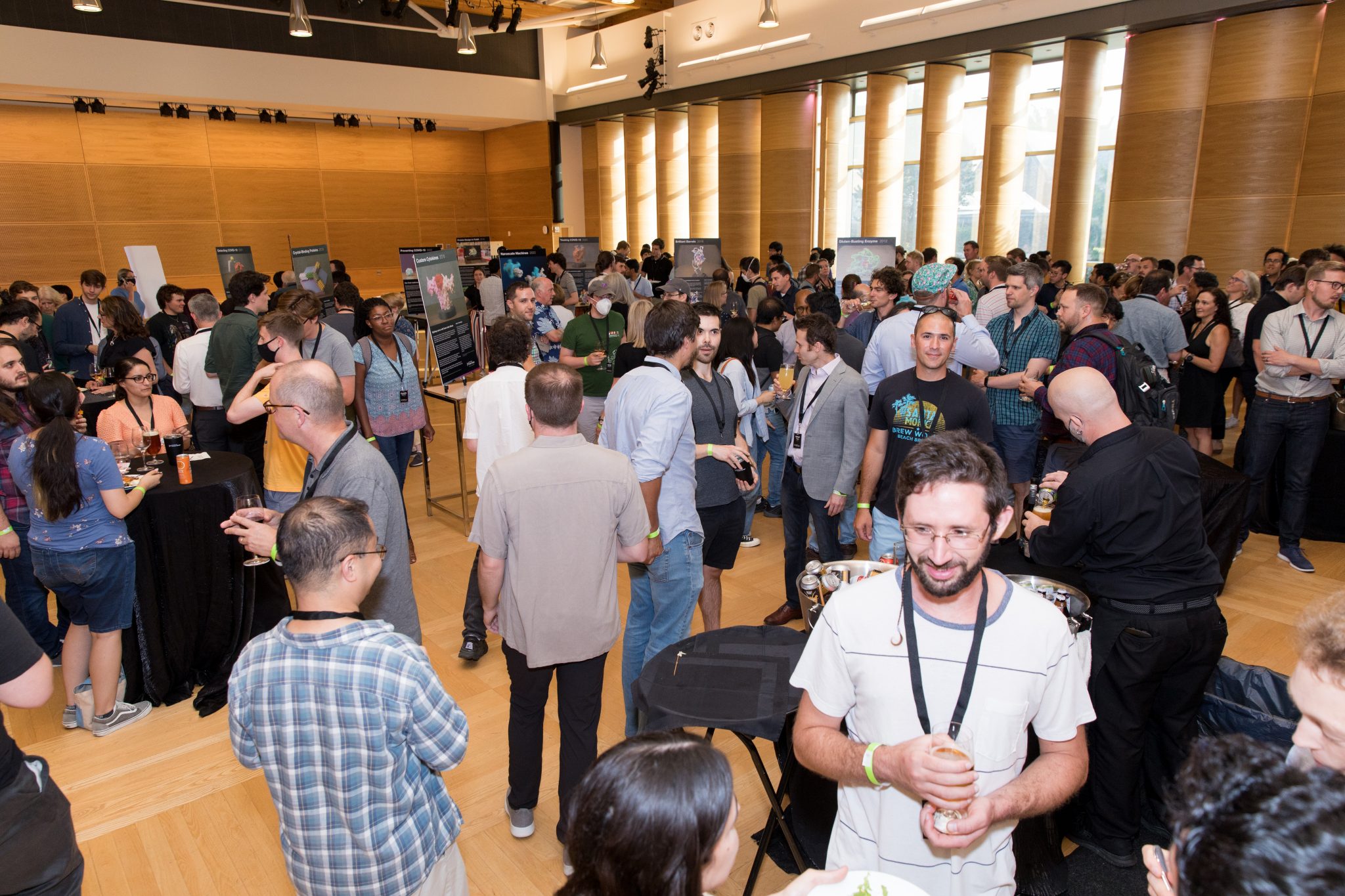


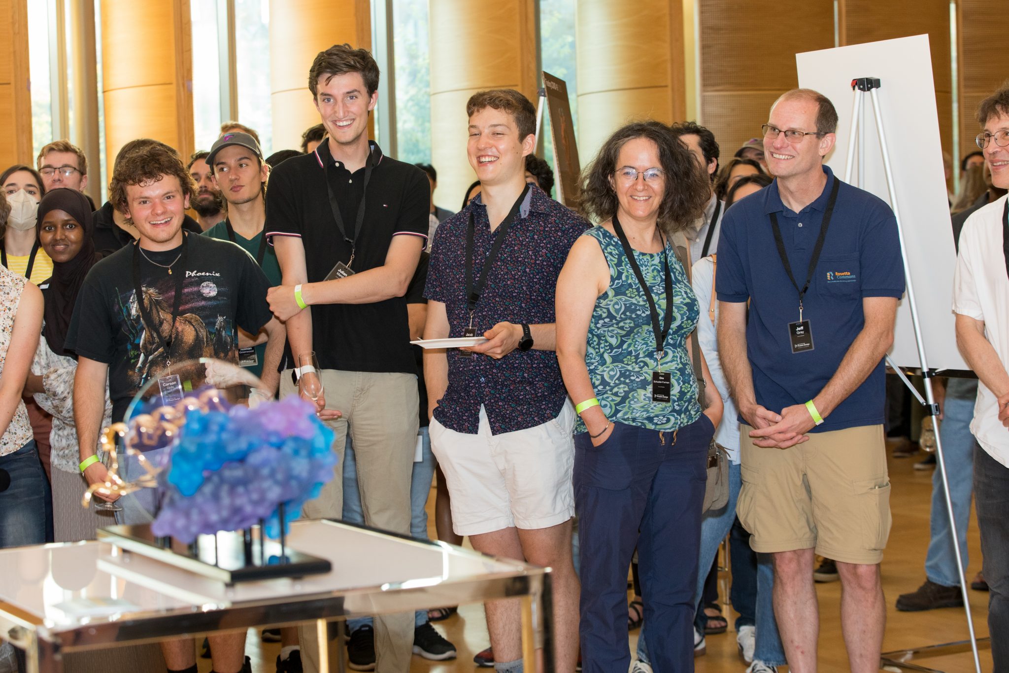
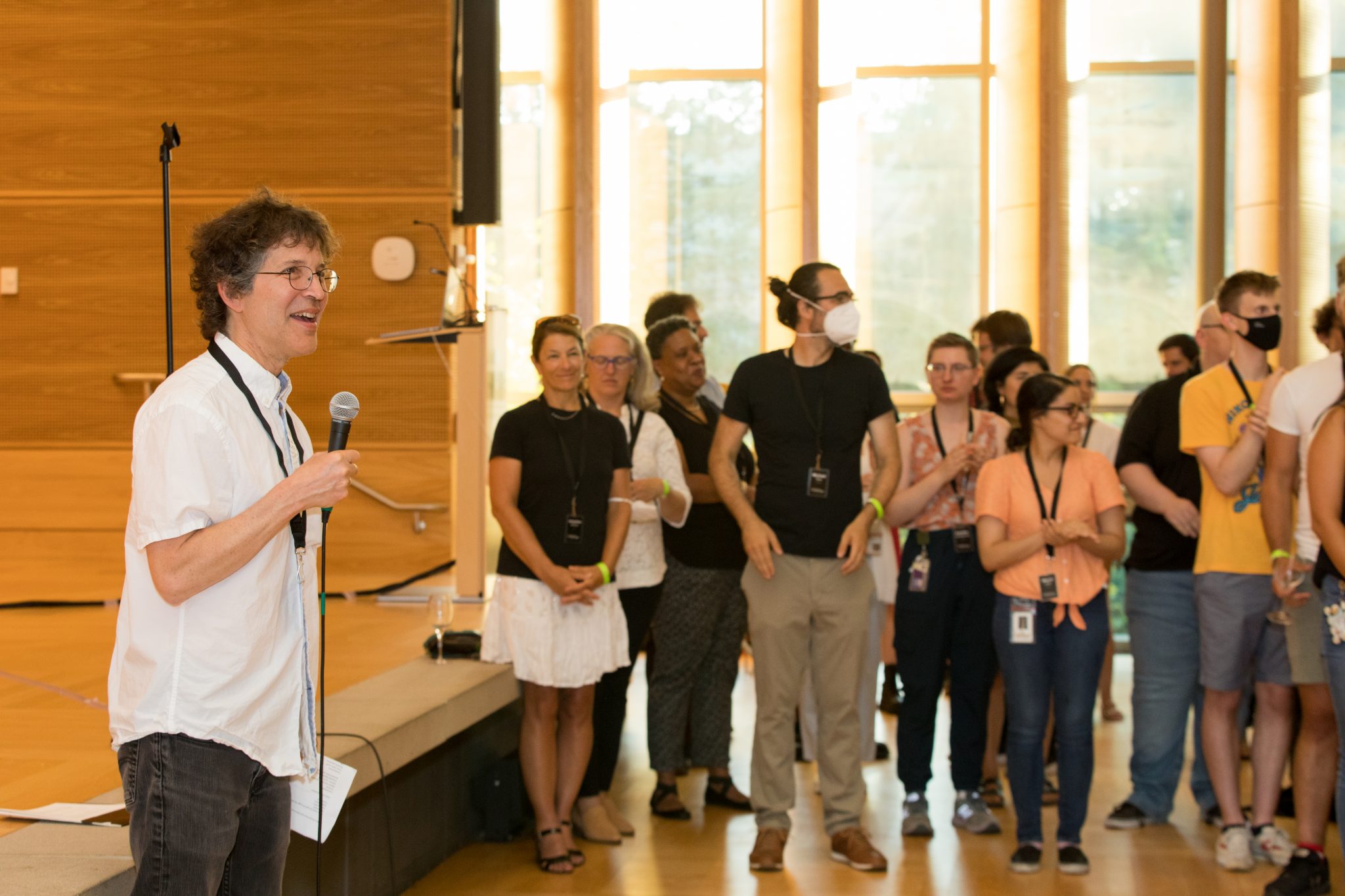
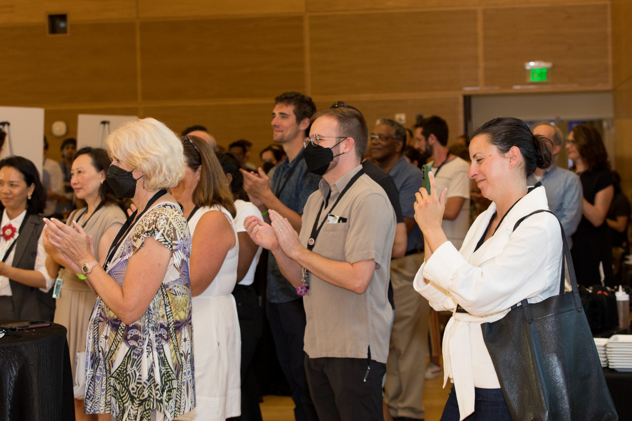
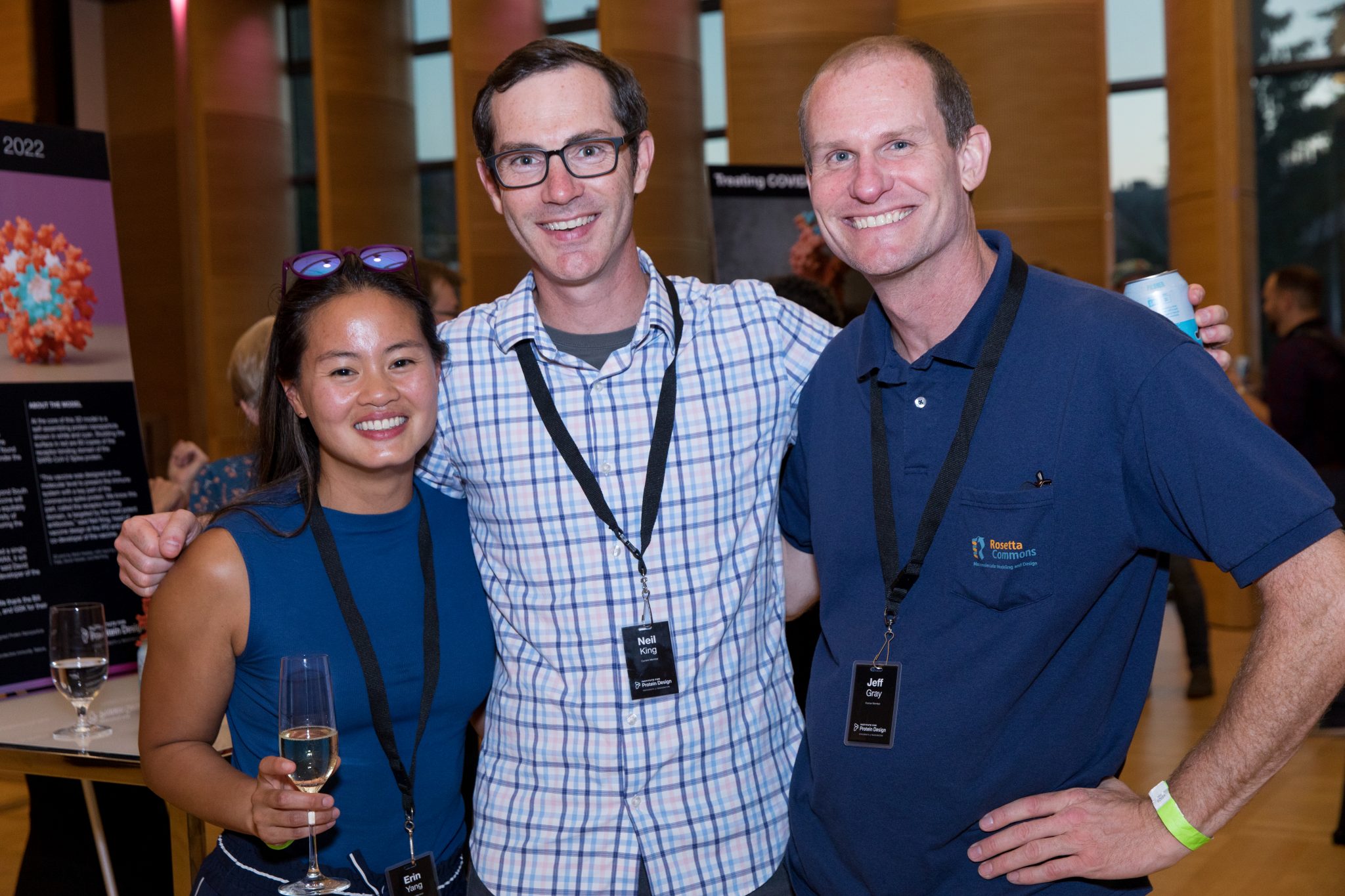
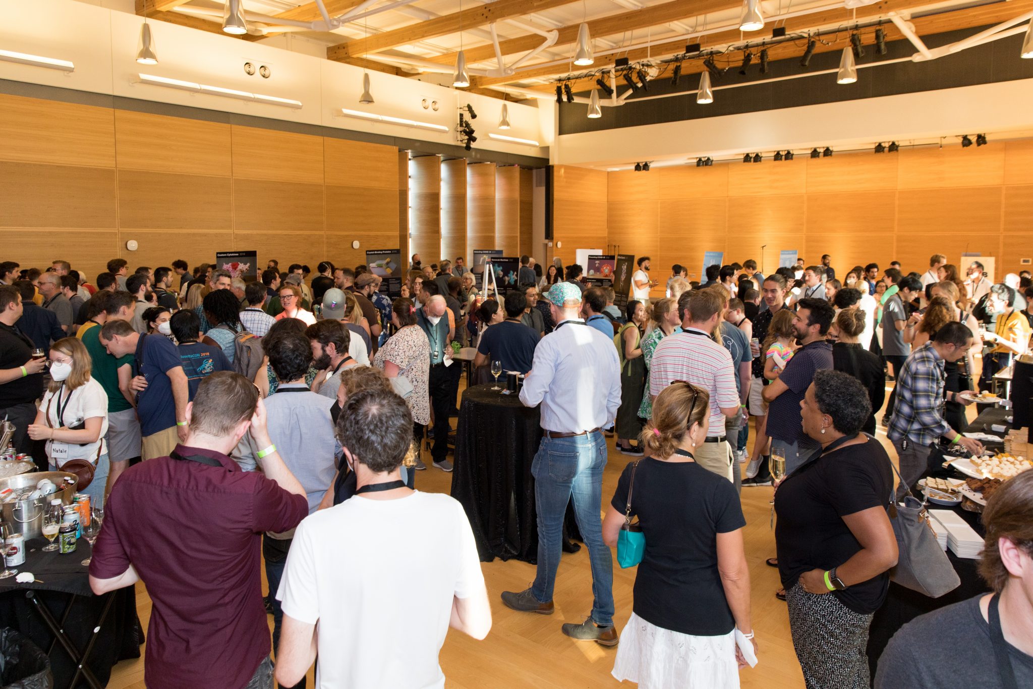
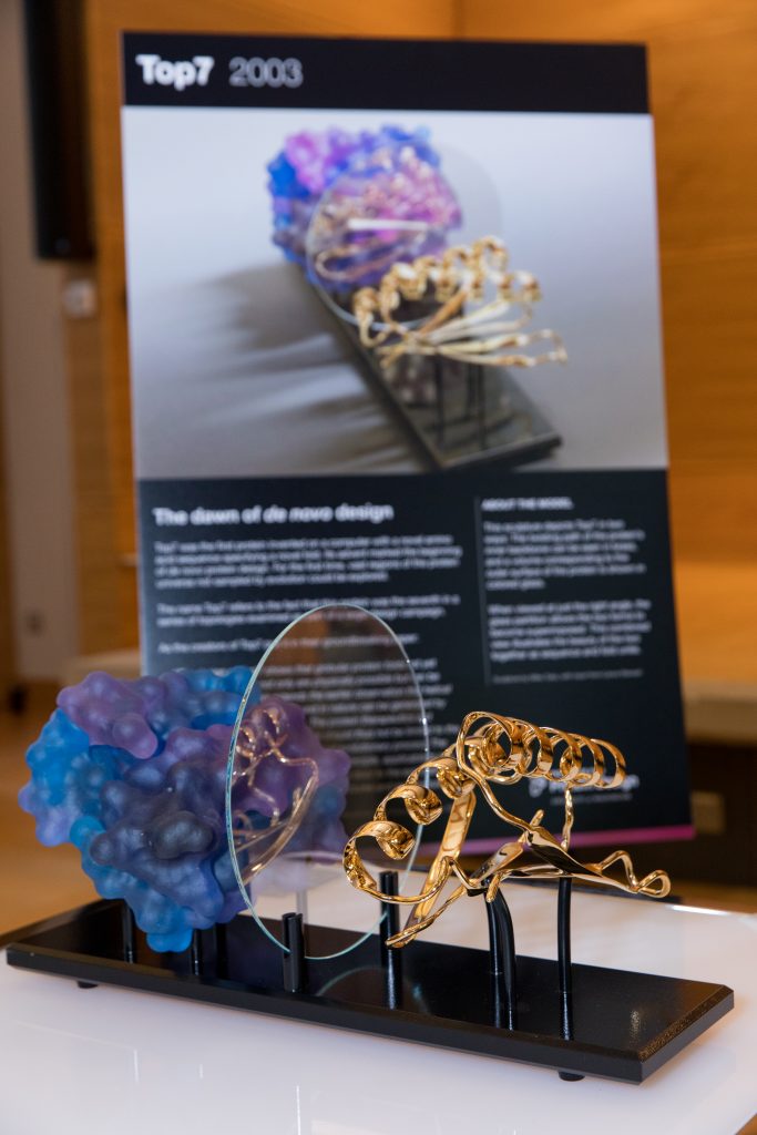 Debuting a new sculpture
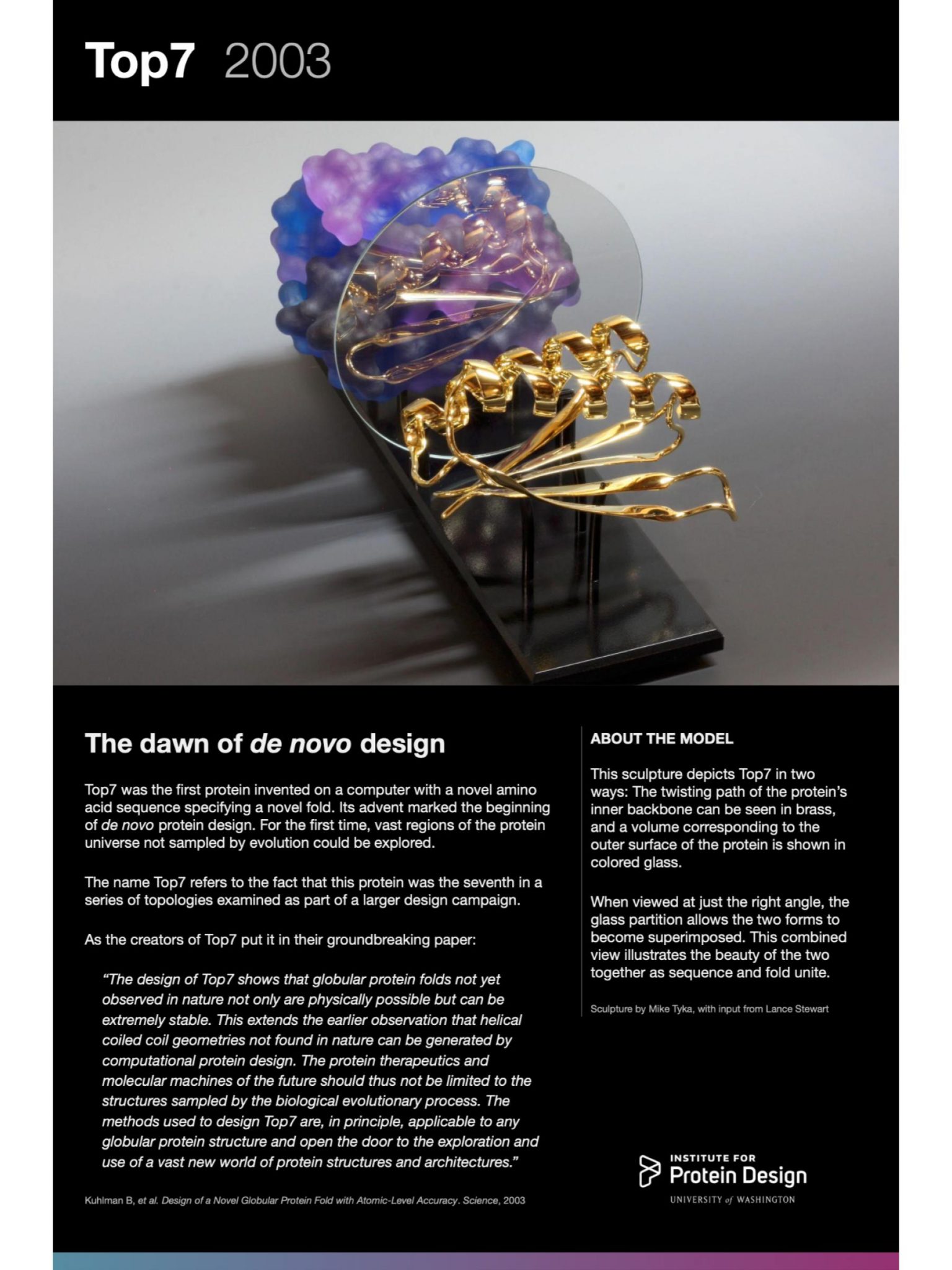
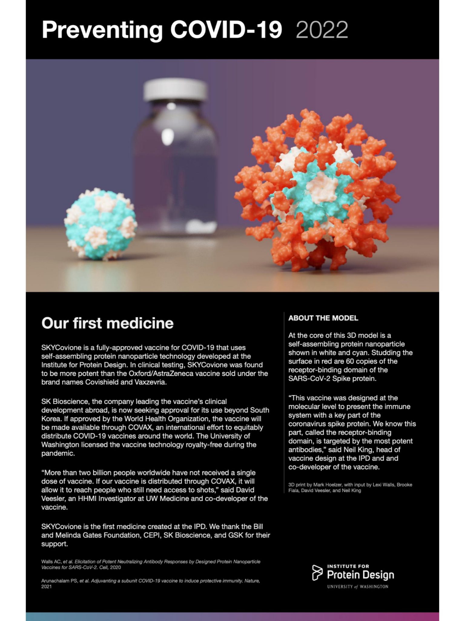
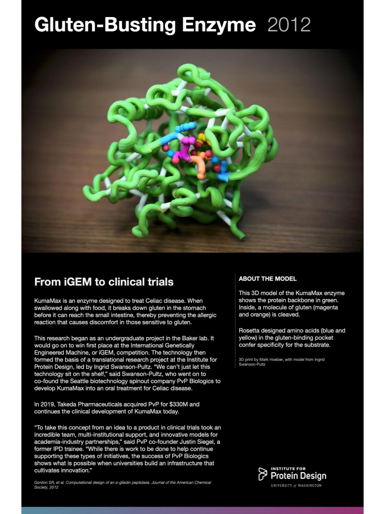
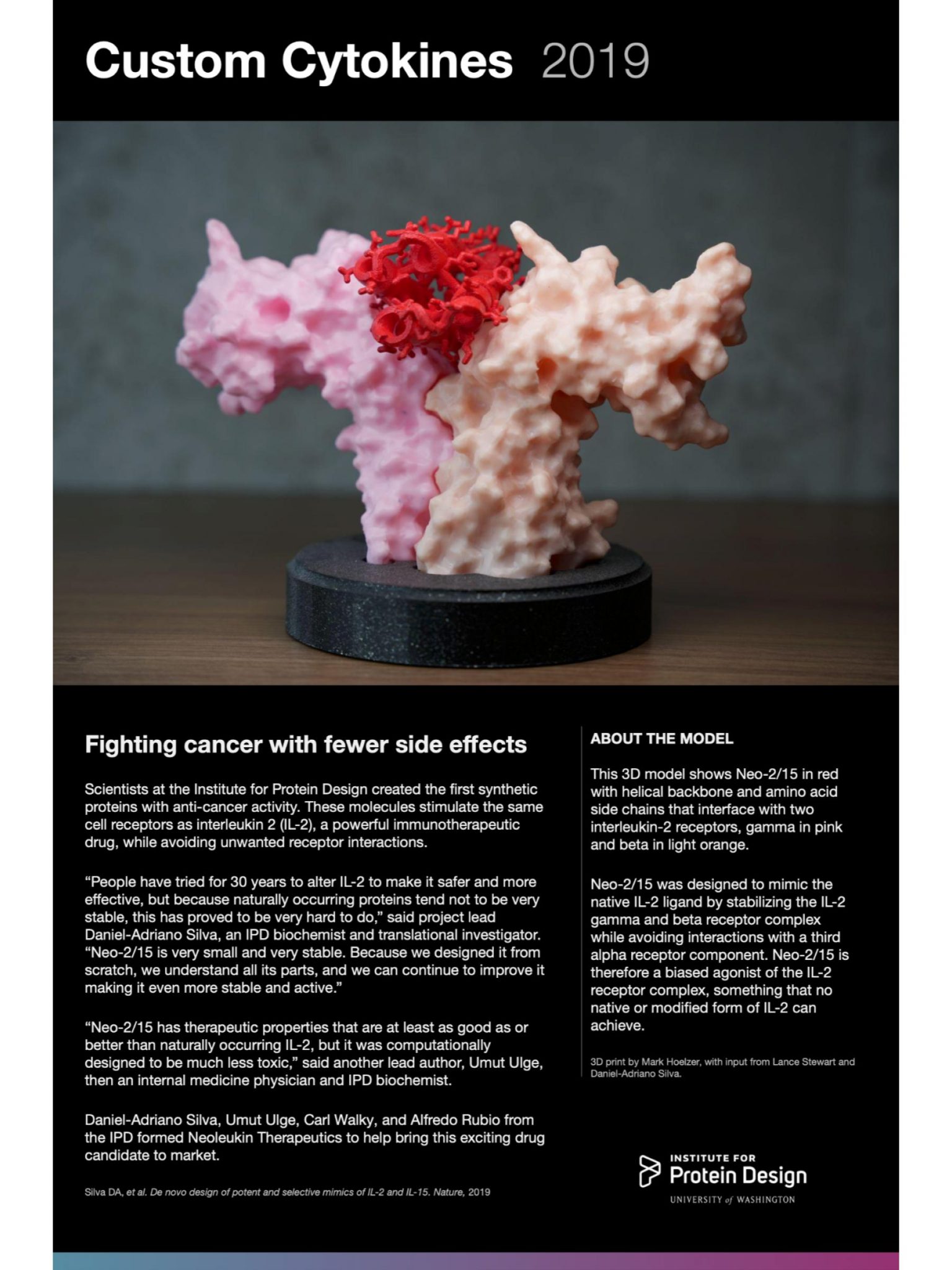
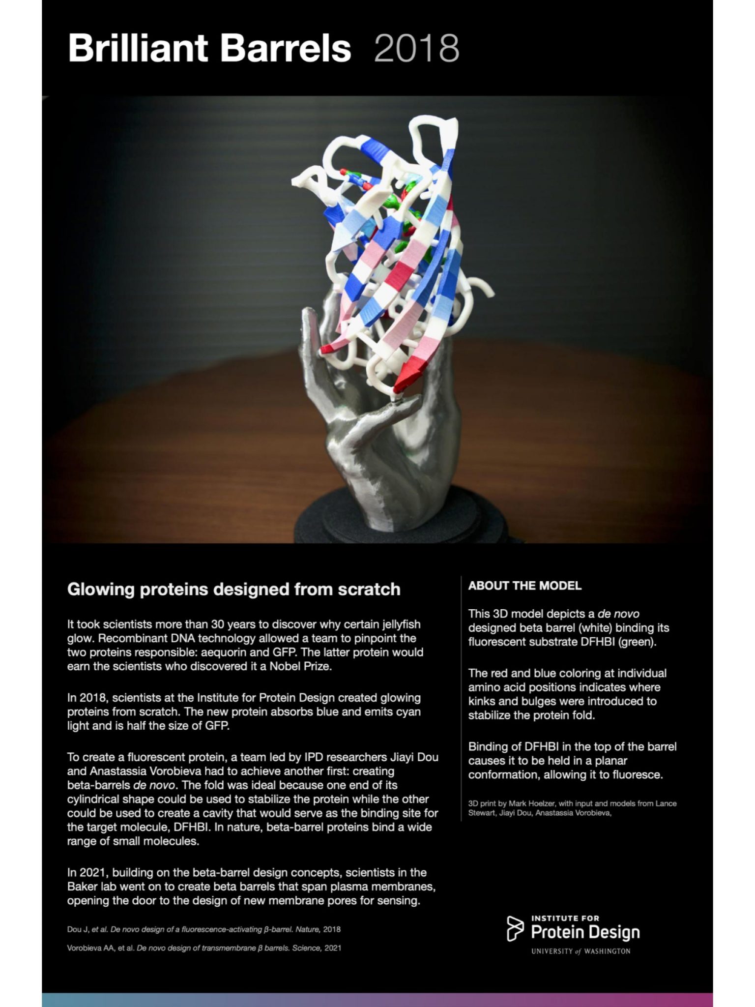
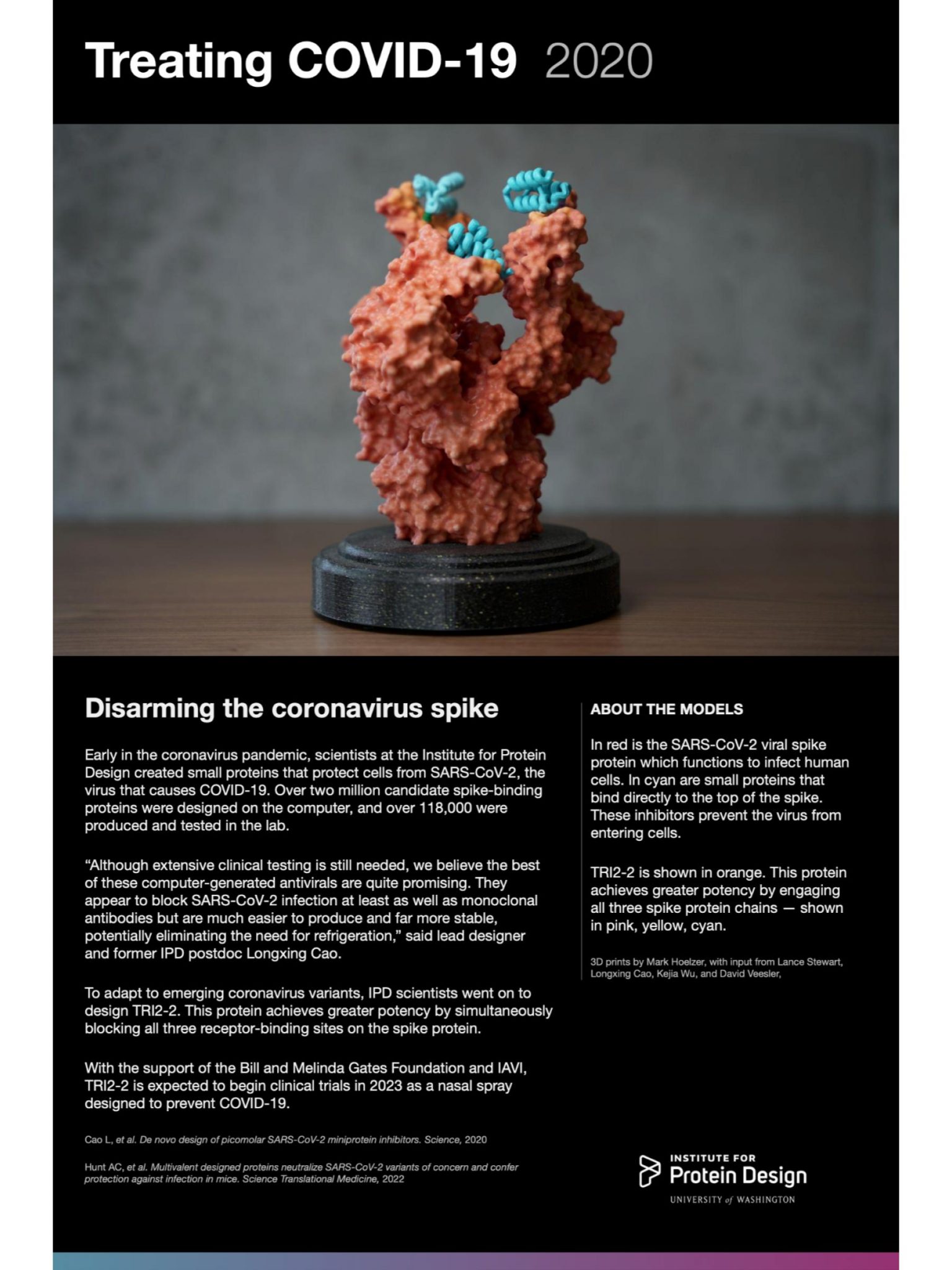
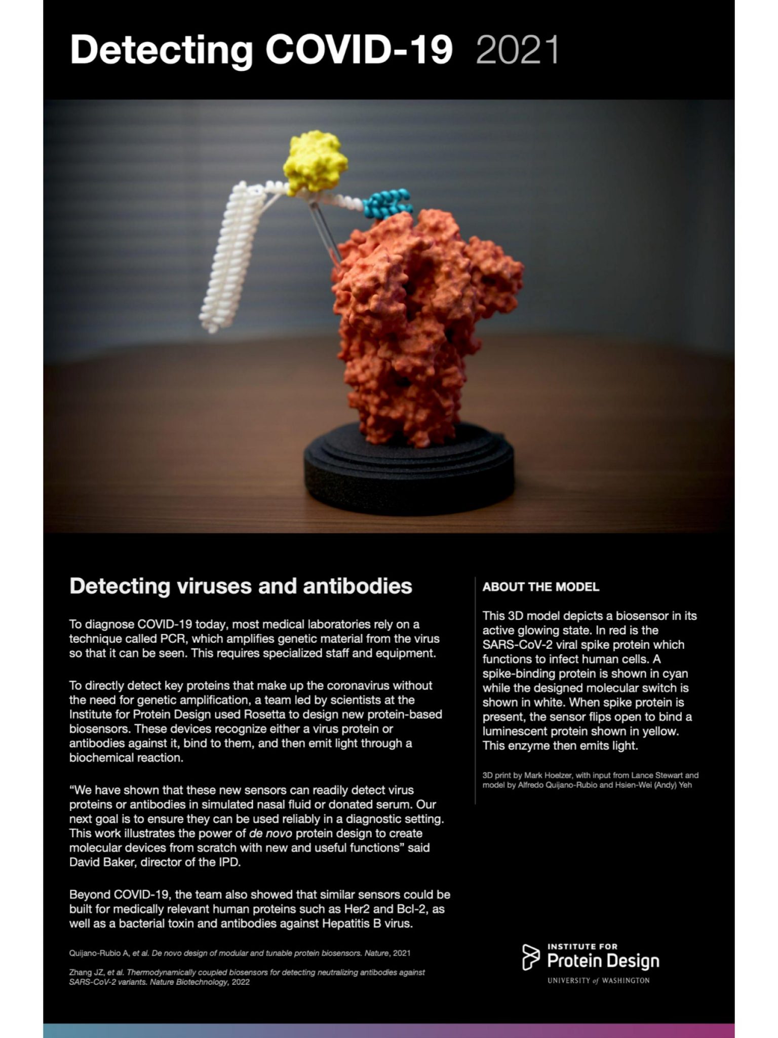
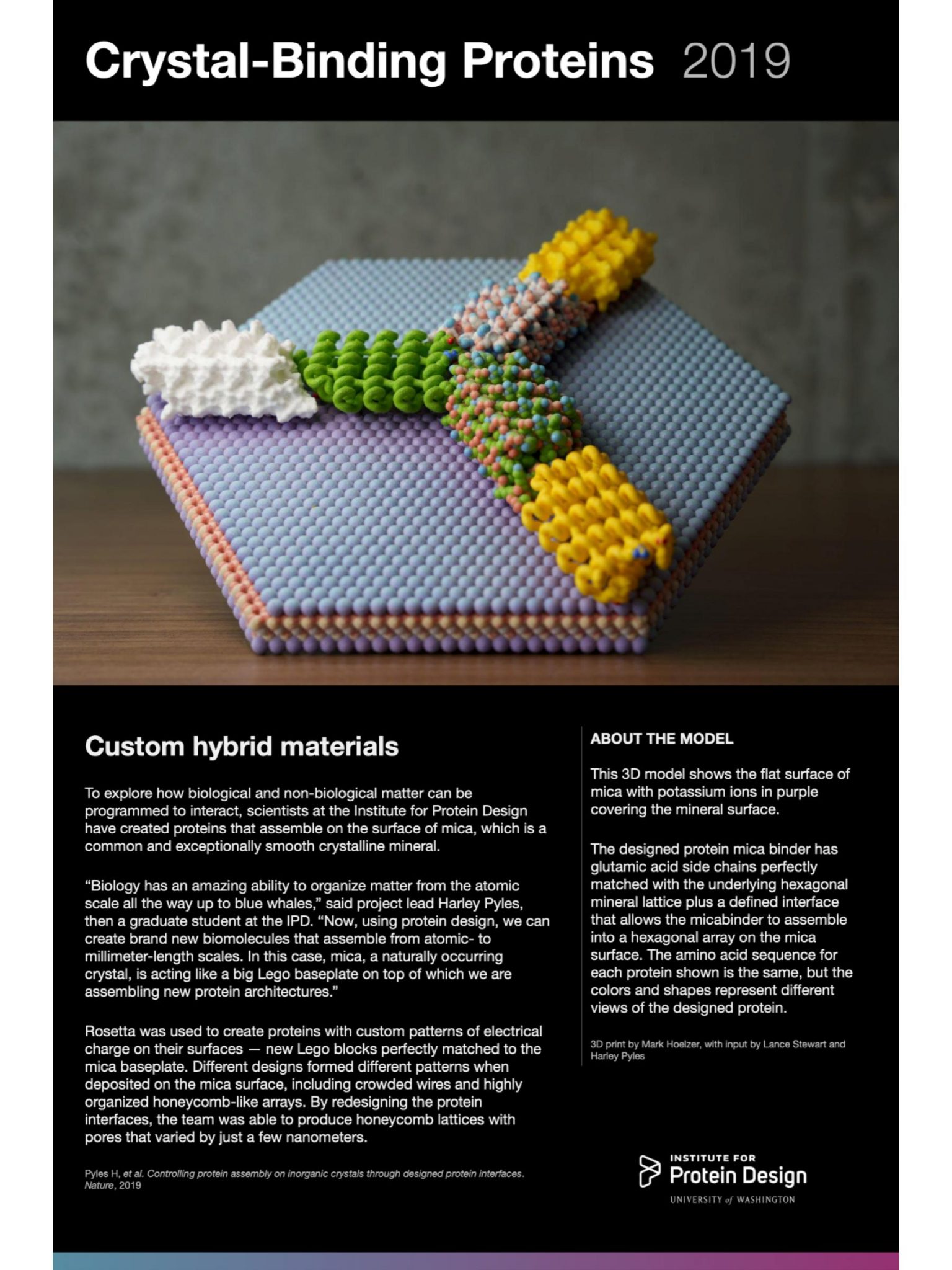
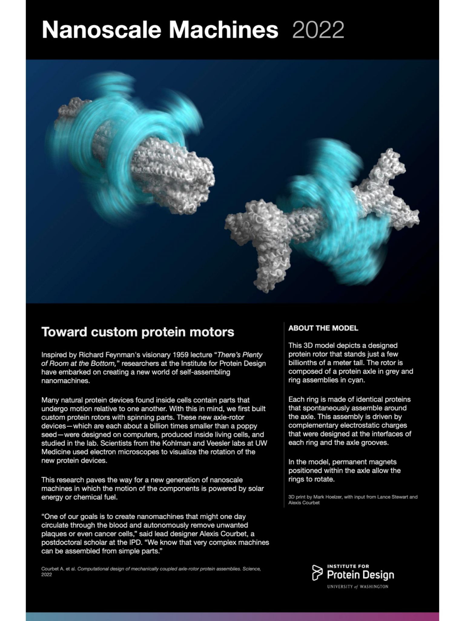
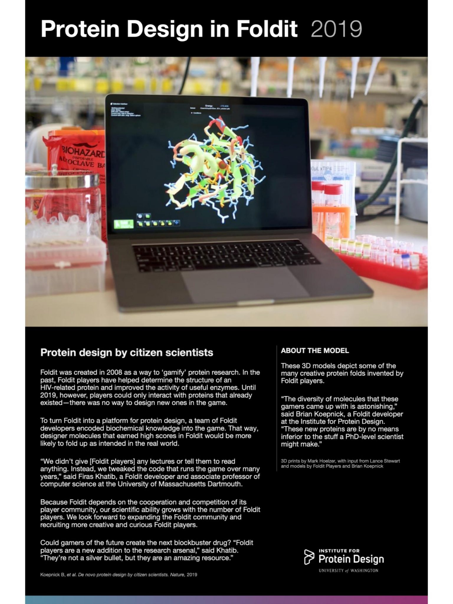
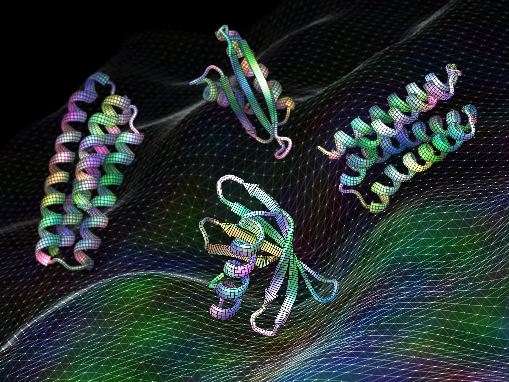
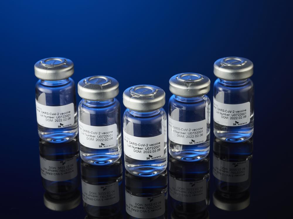


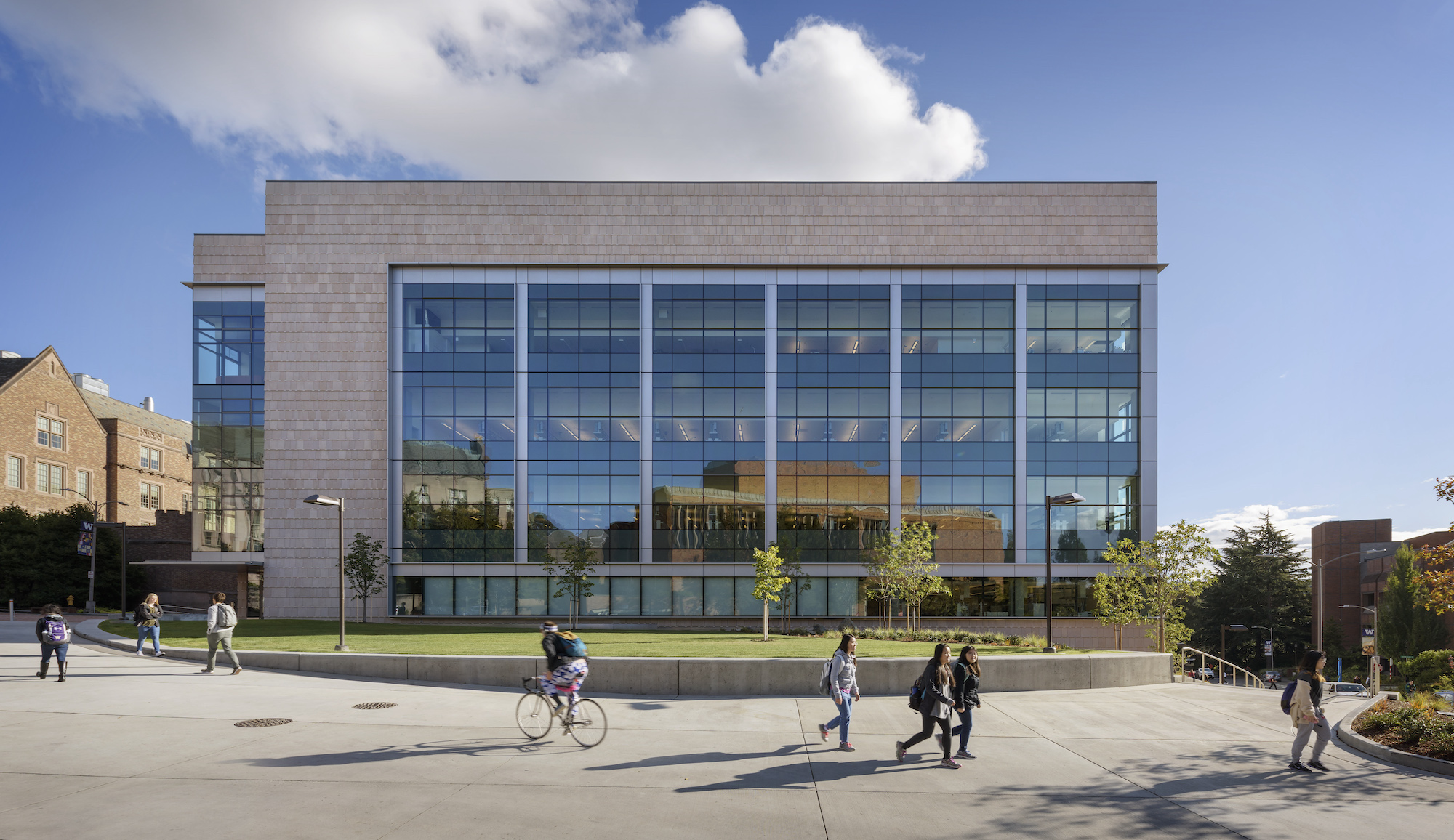

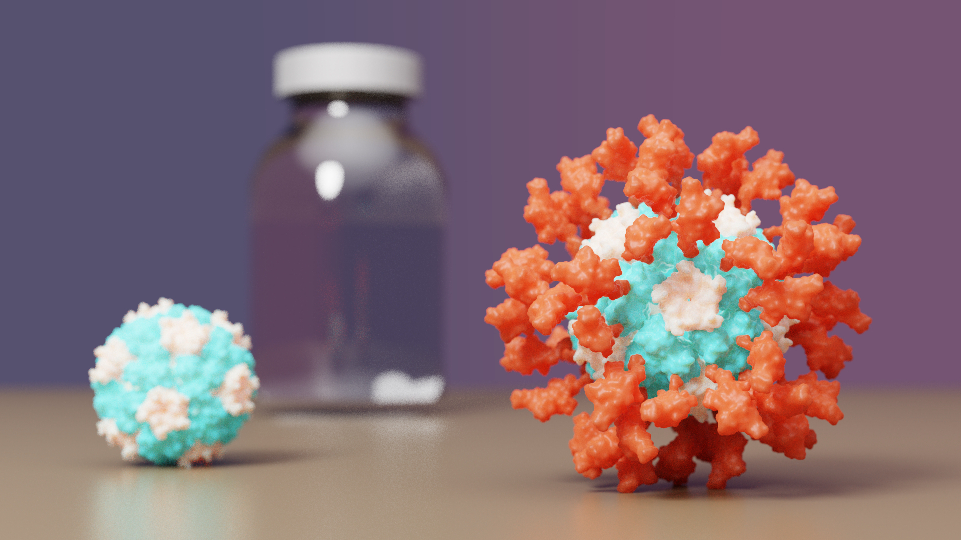

 “This vaccine was designed at the molecular level to present the immune system with a key part of the coronavirus spike protein. We know this part, called the receptor-binding domain, is targeted by the most potent antibodies,” said
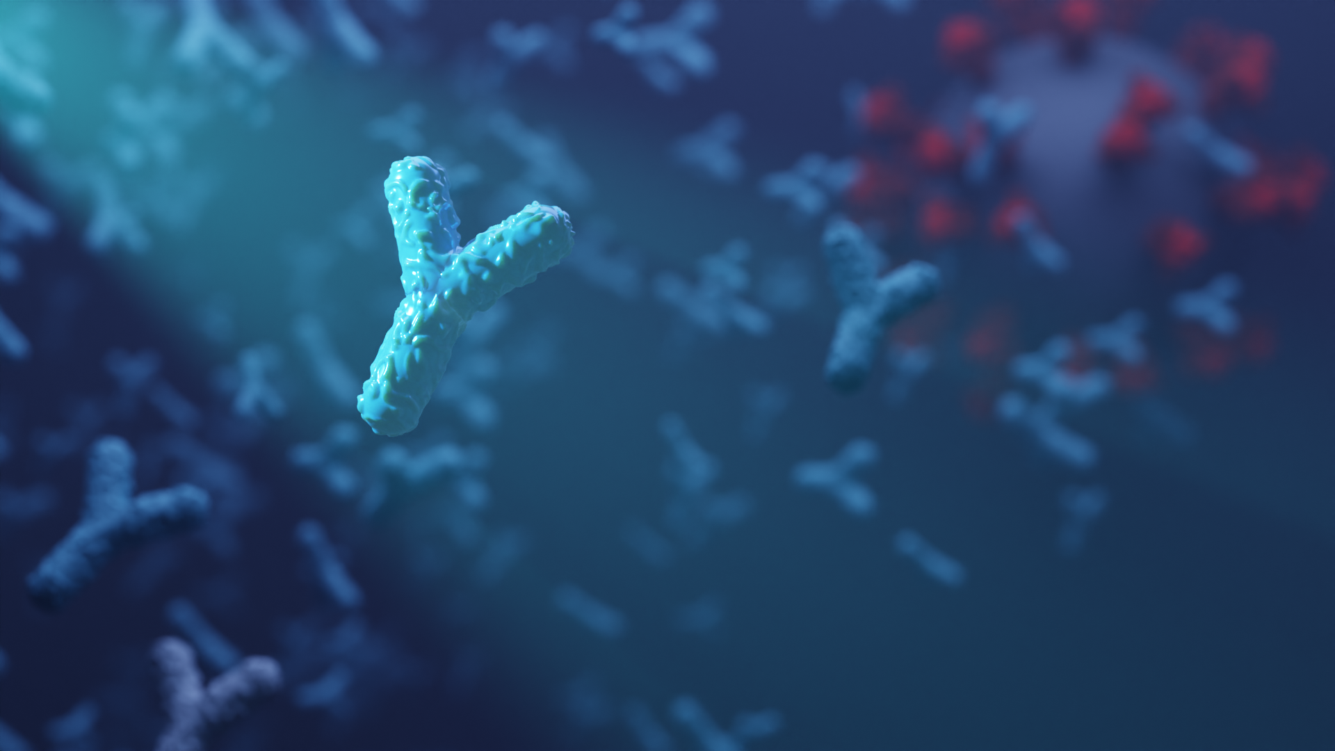
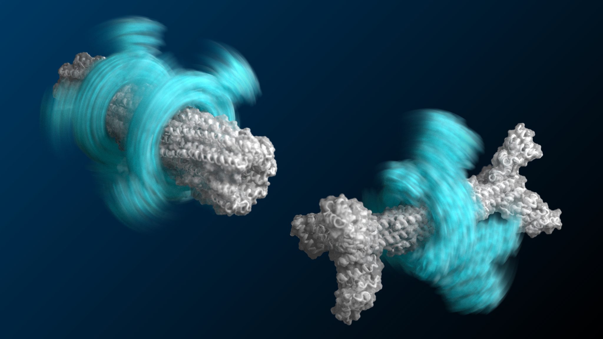
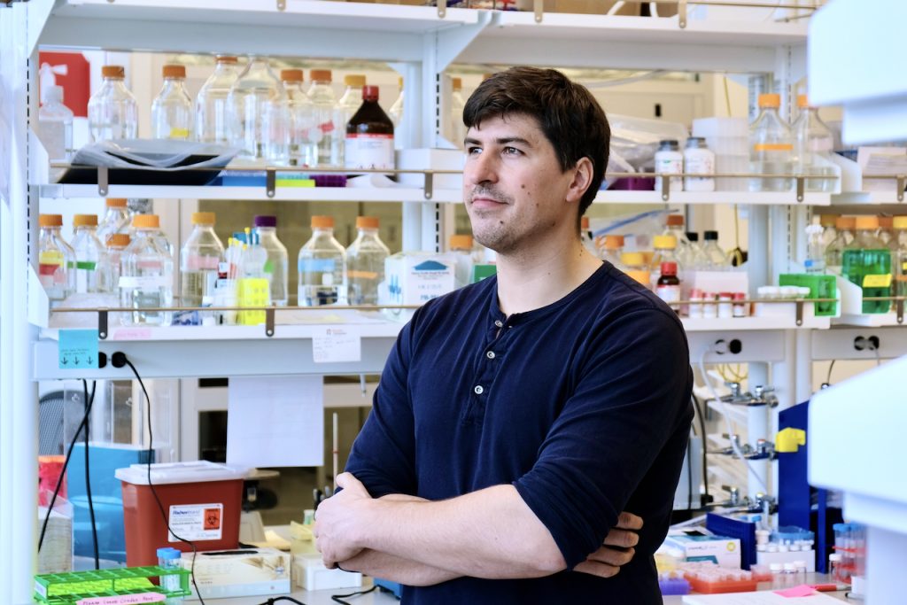
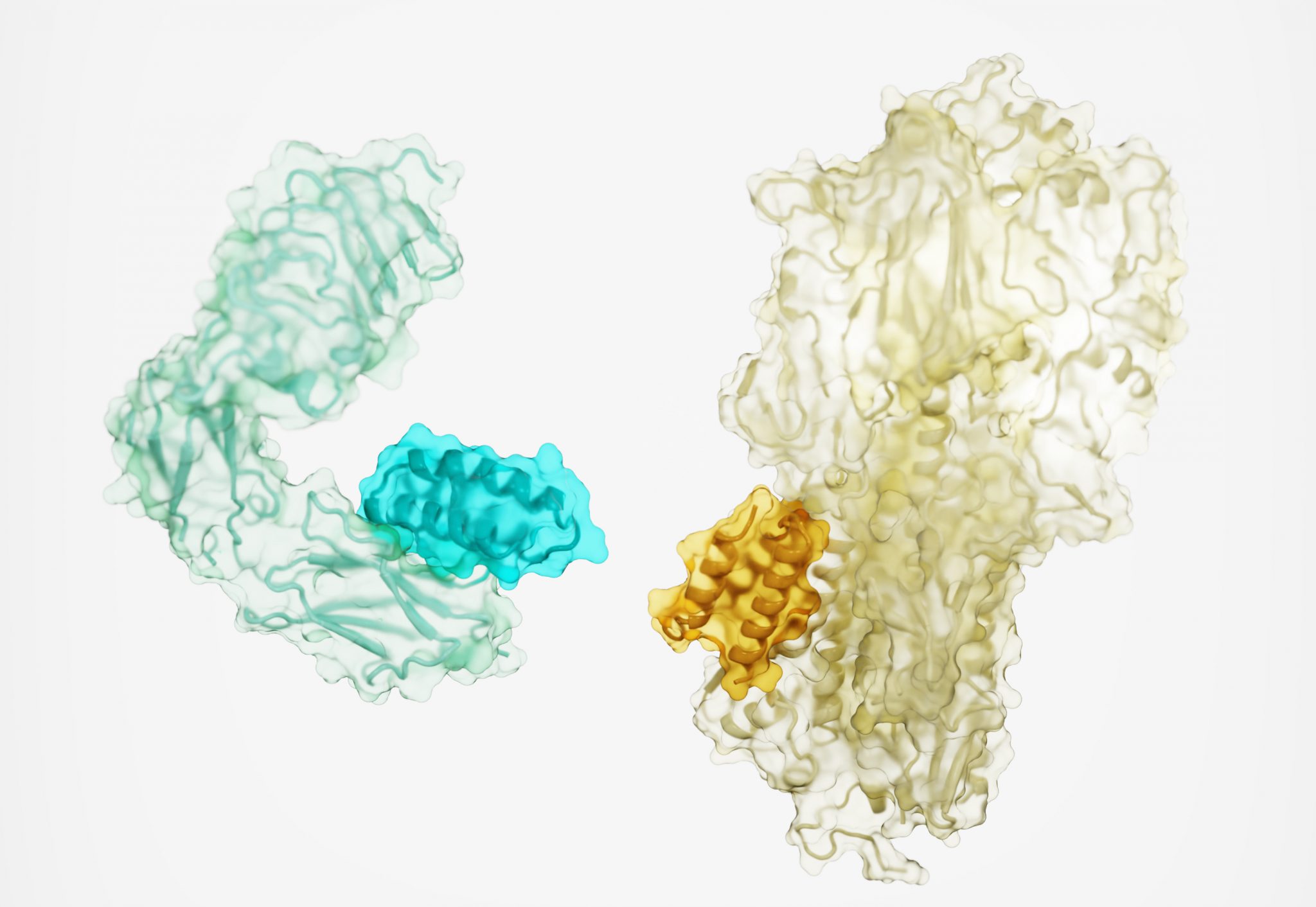
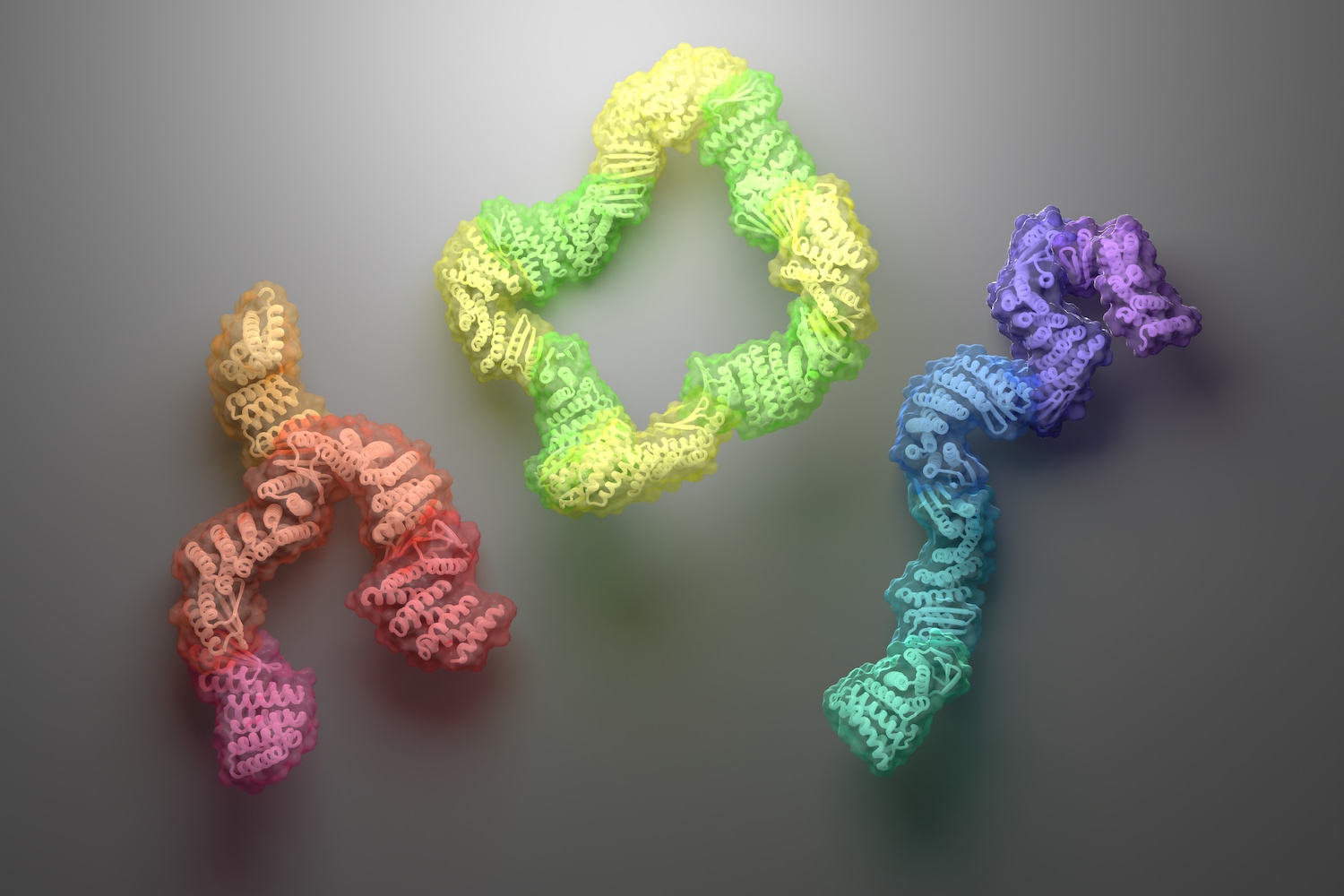

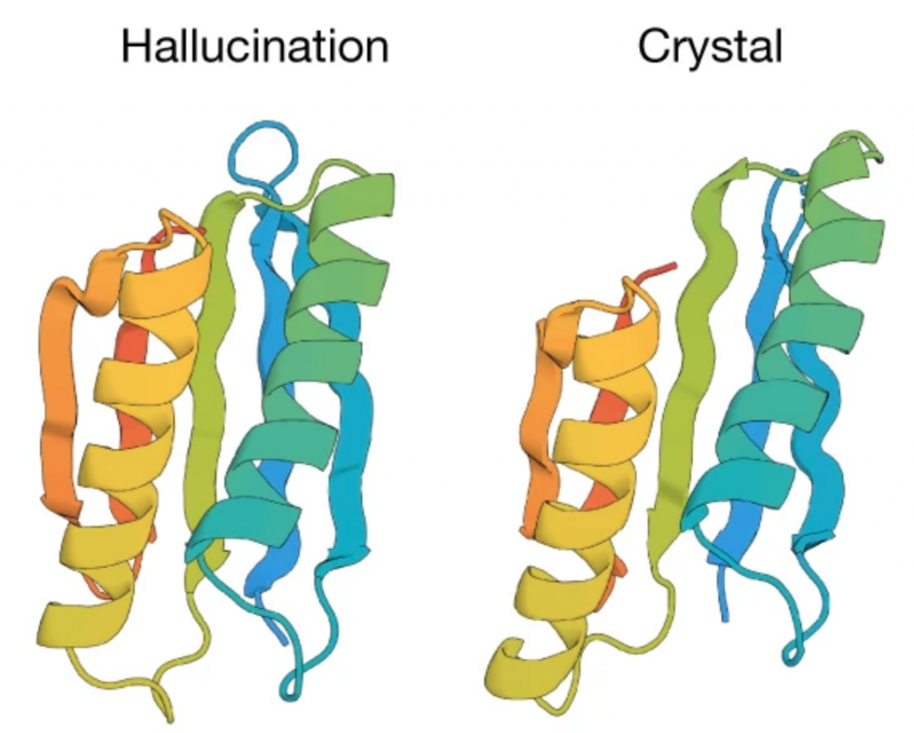
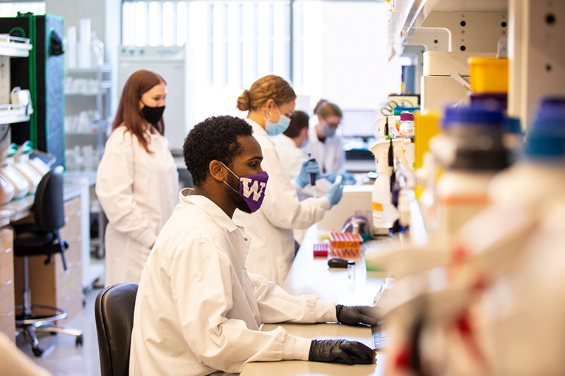
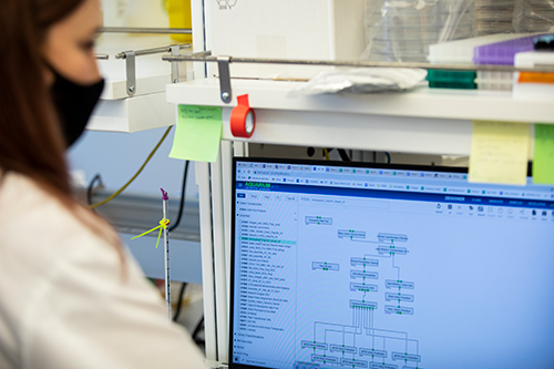
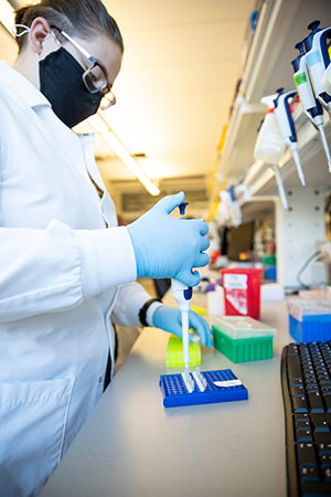
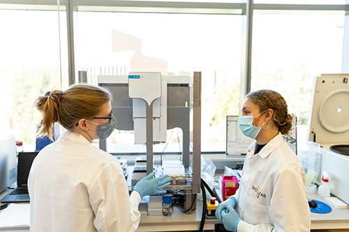
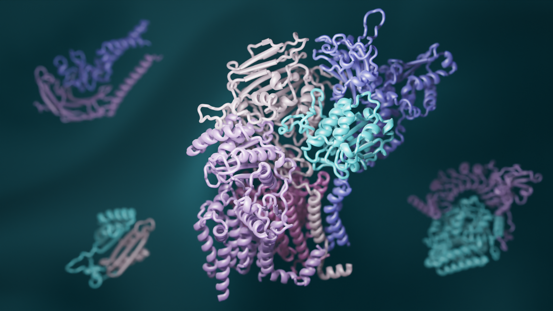
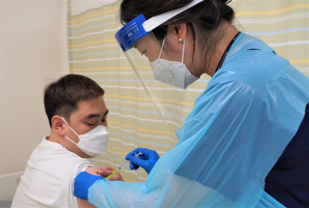
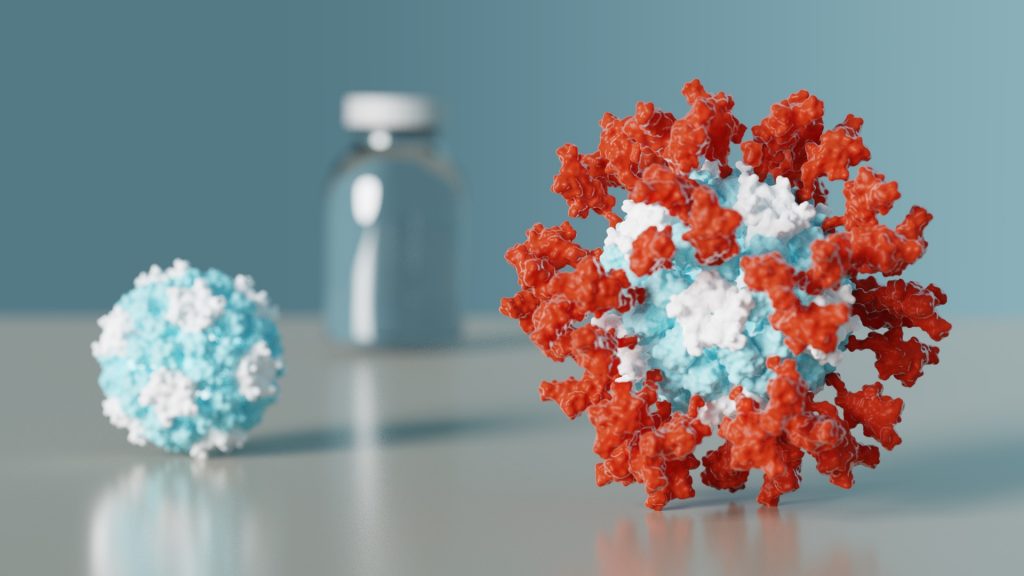
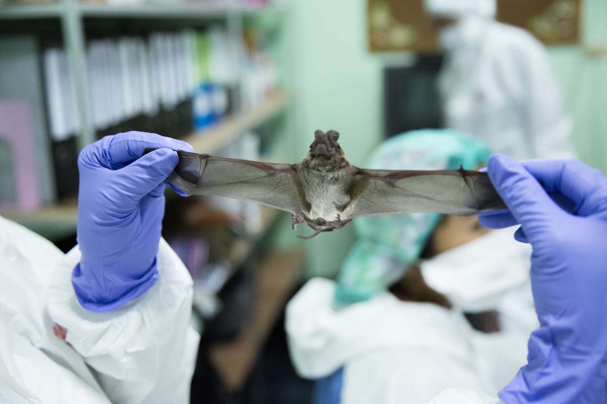

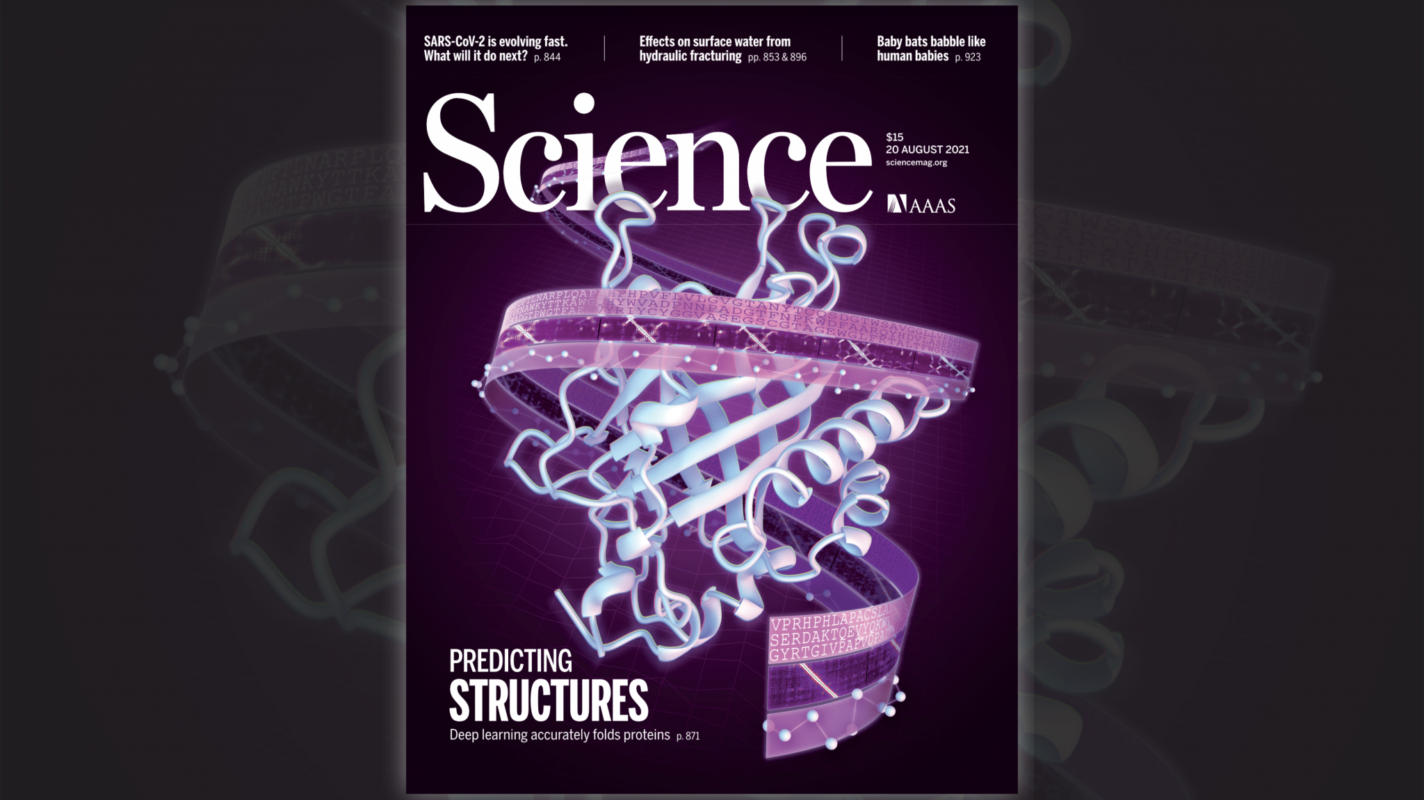

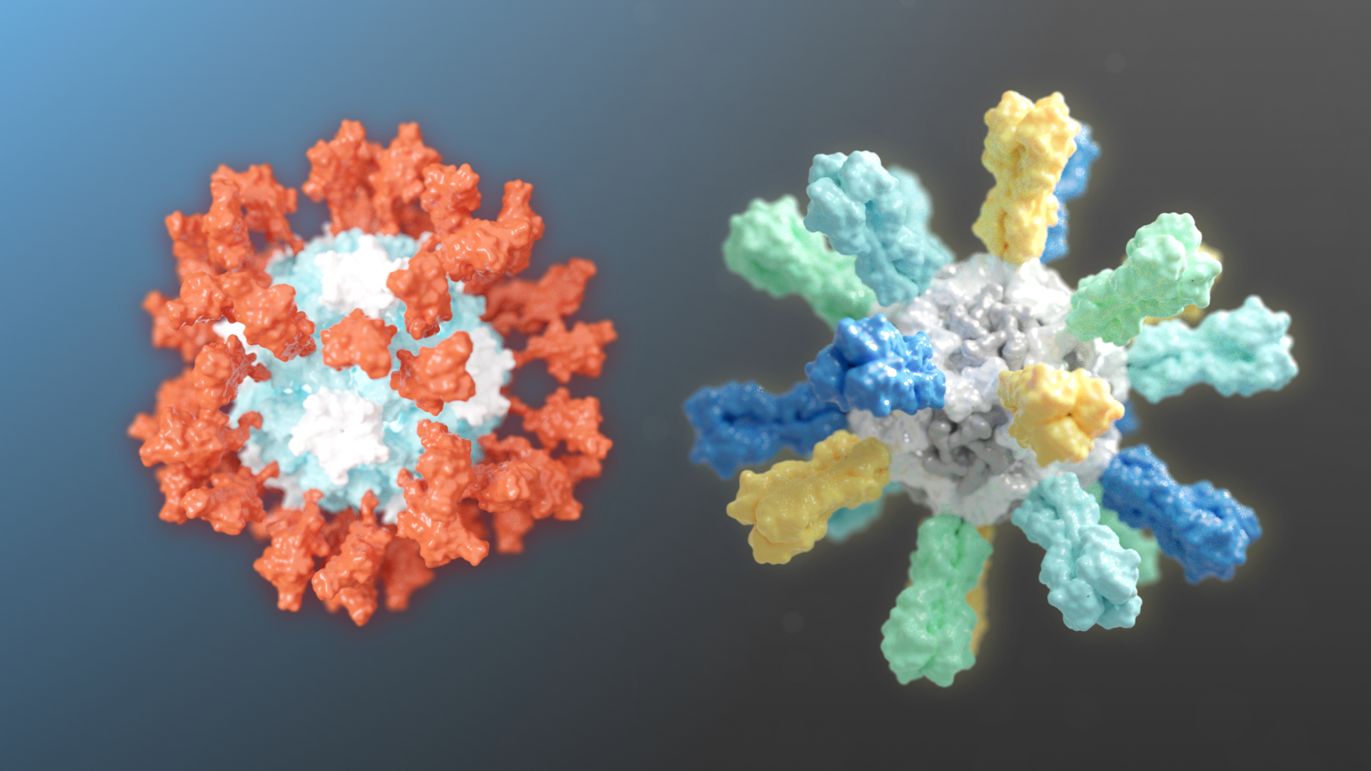
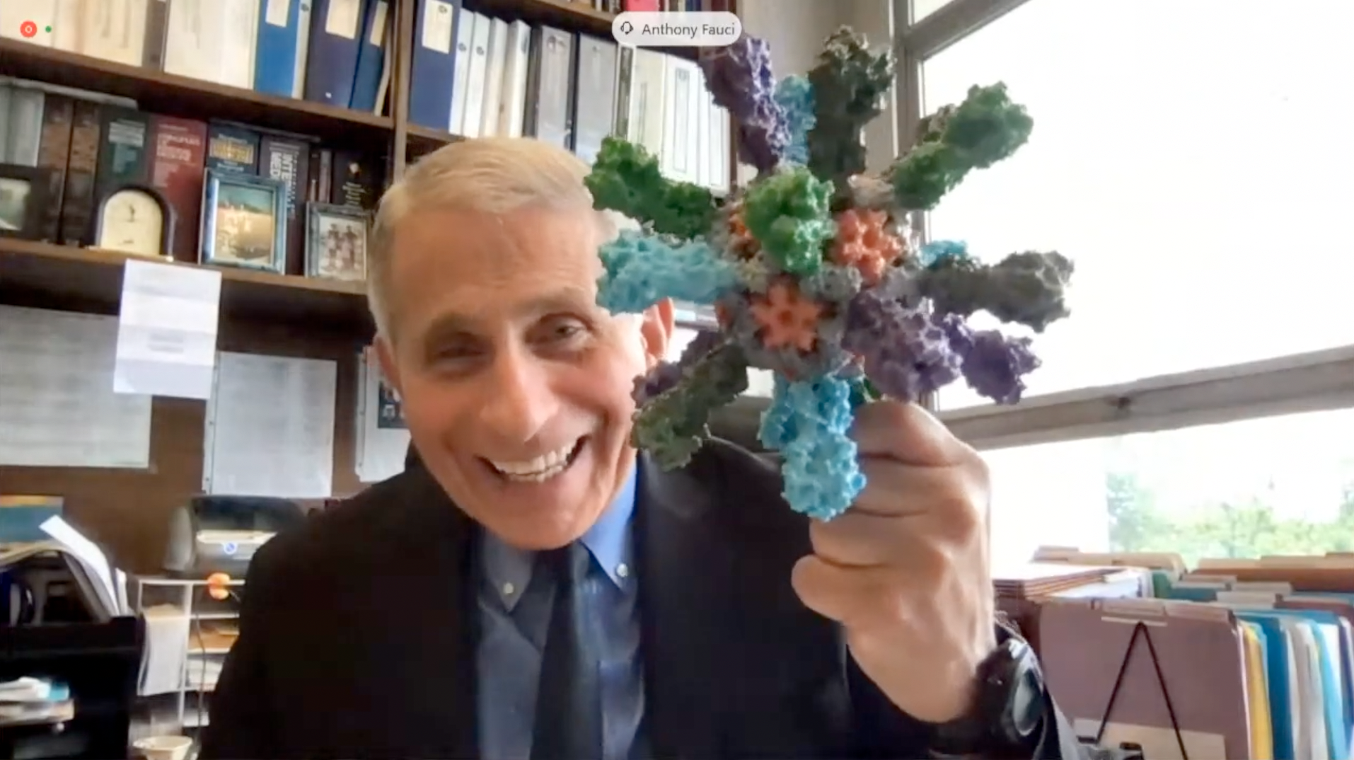
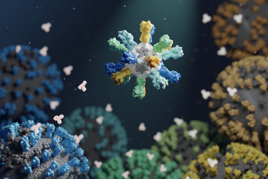
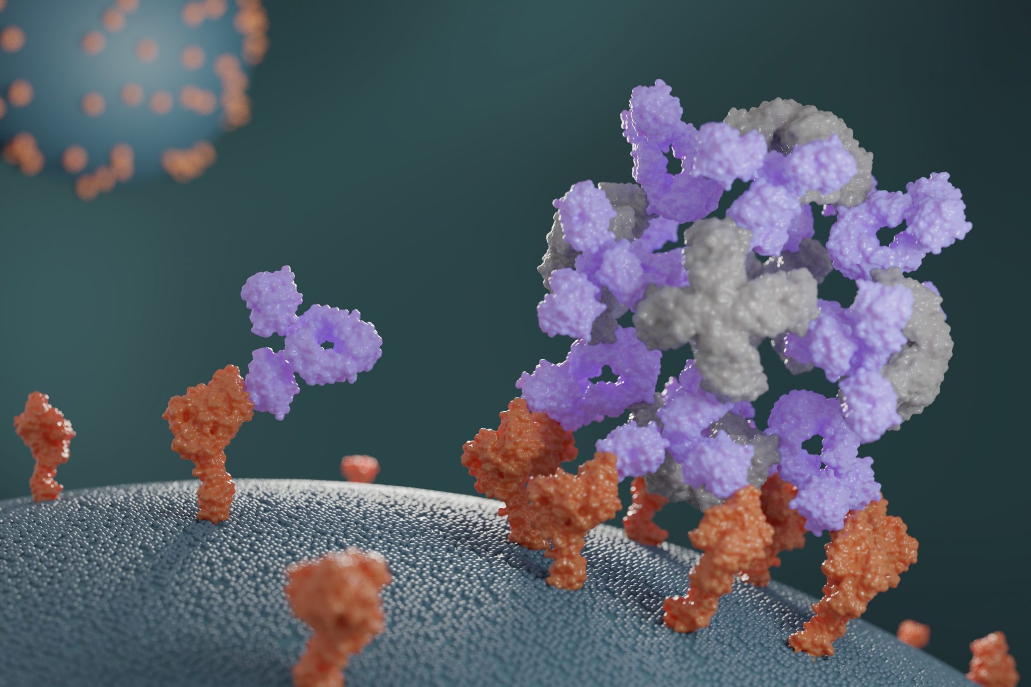
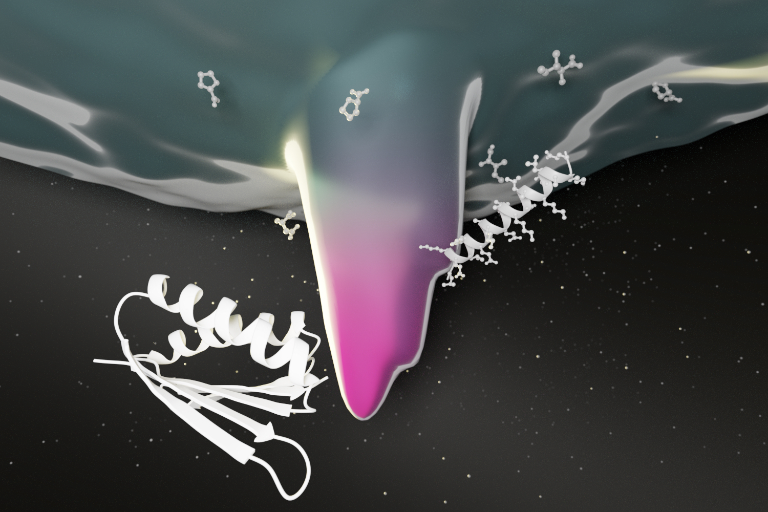
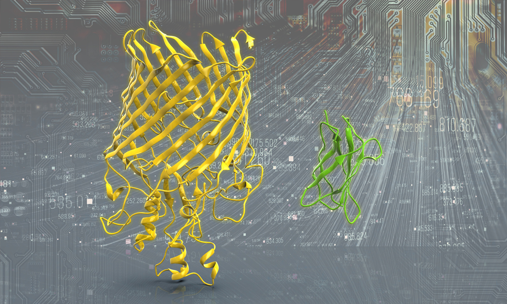

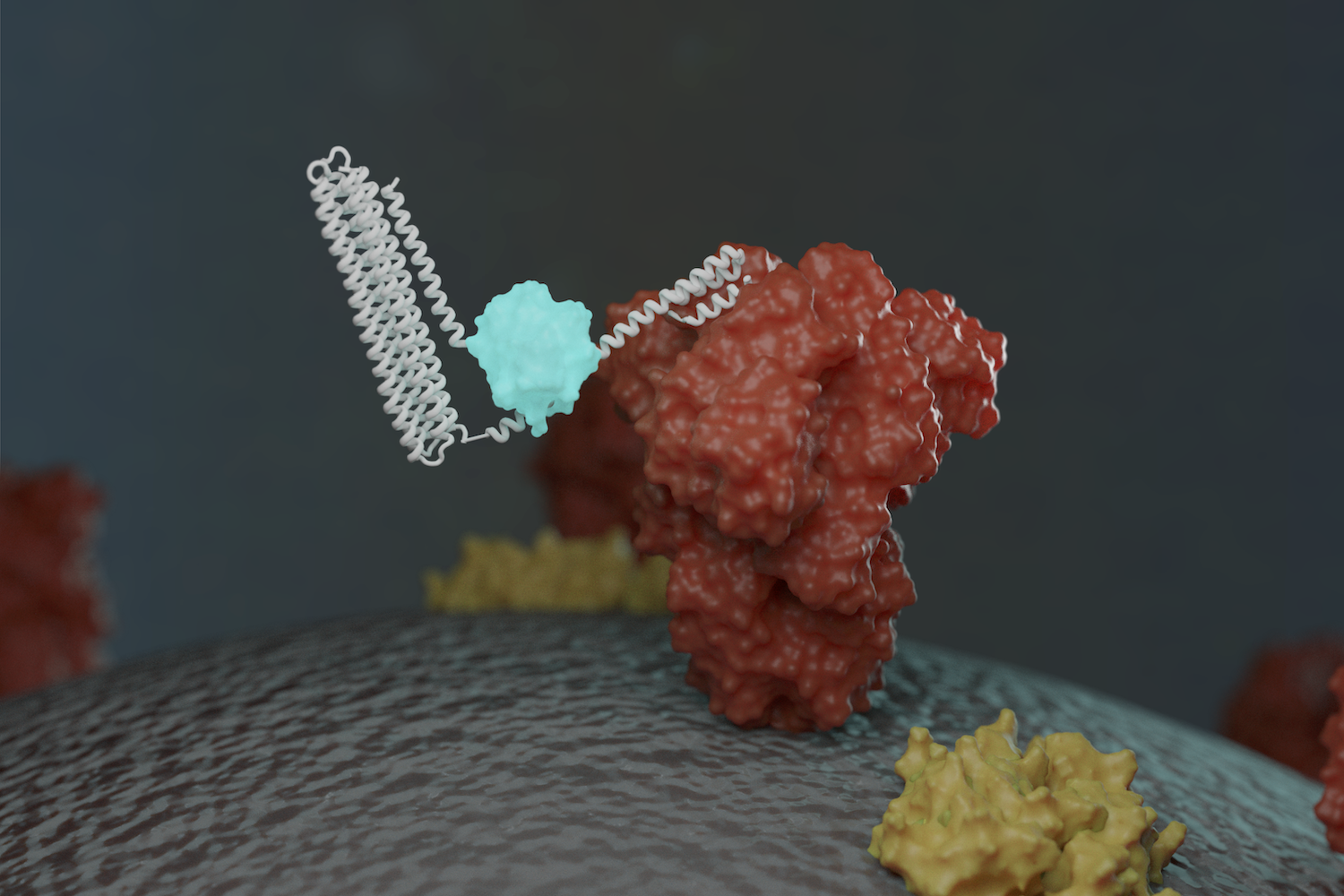

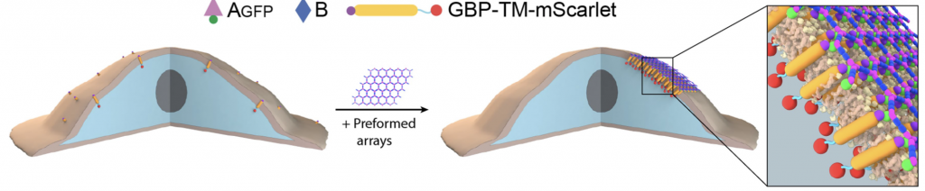

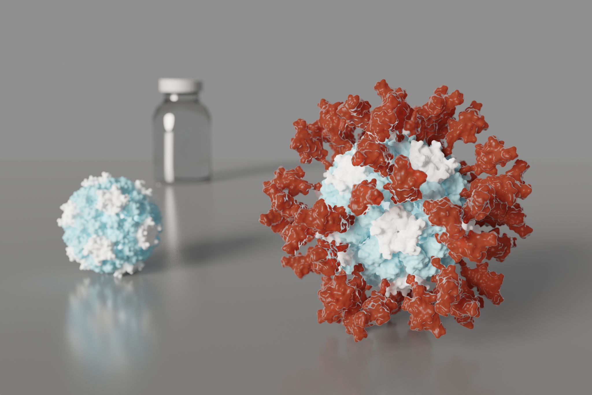

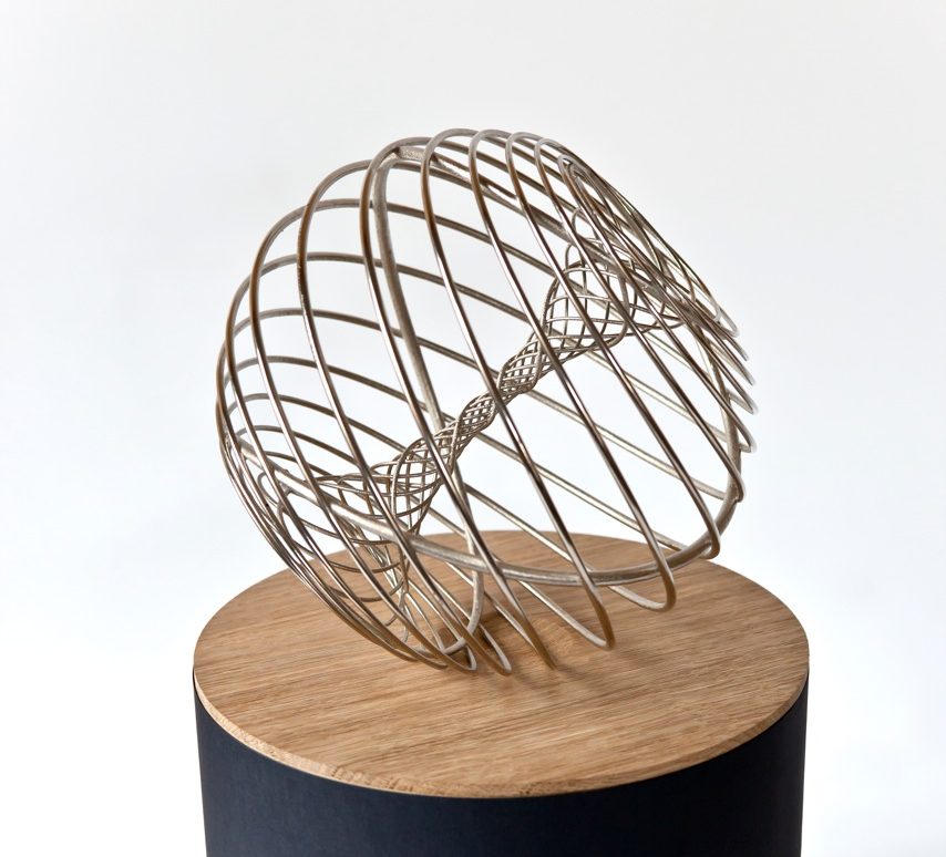
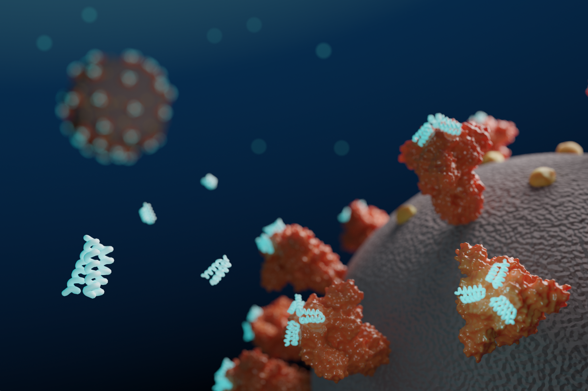
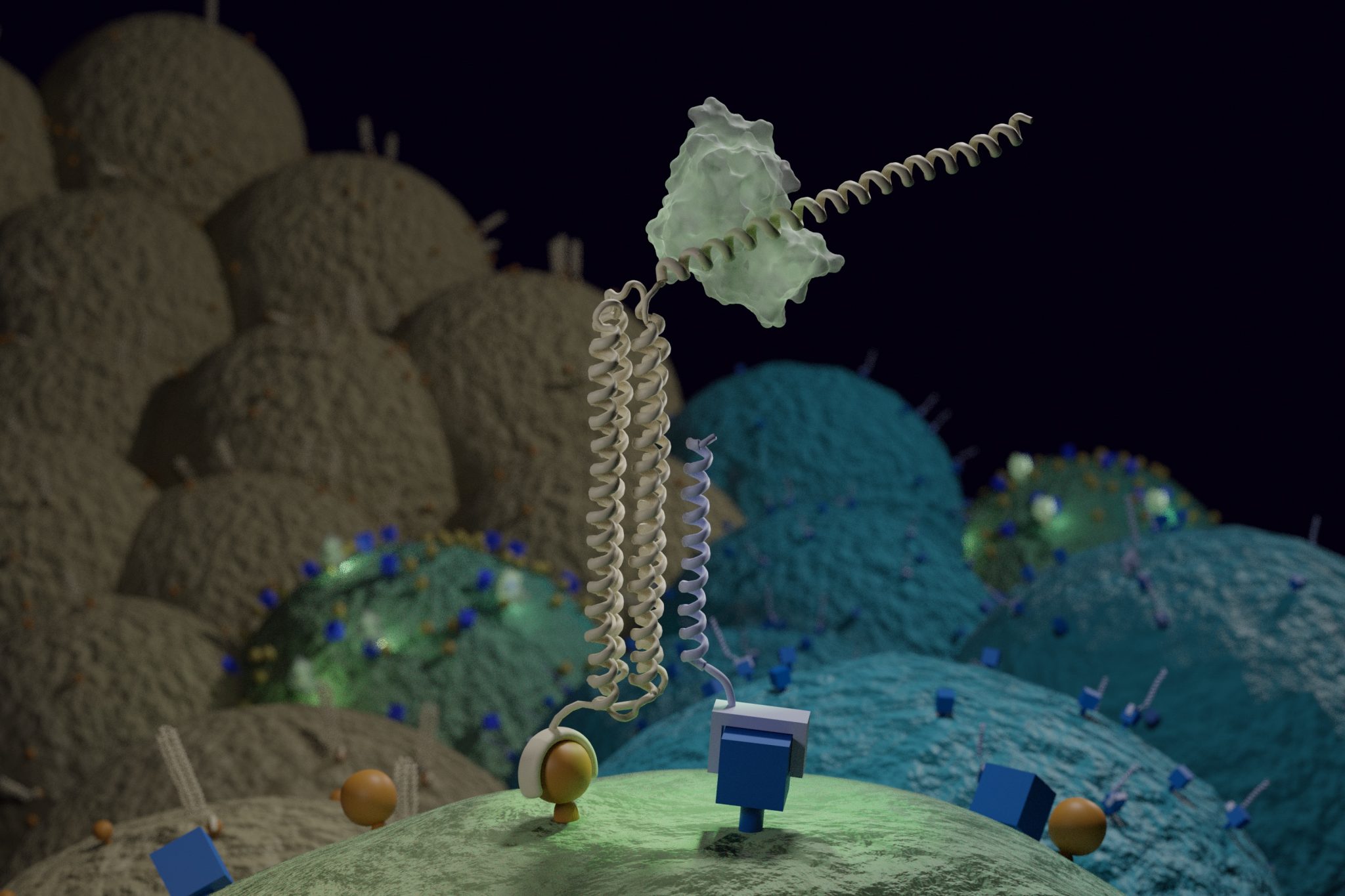

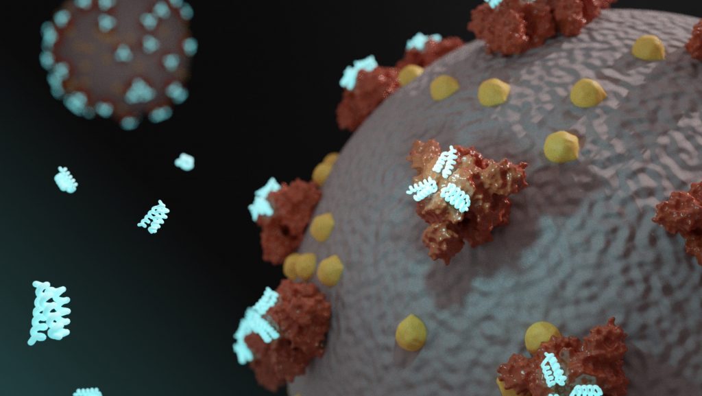
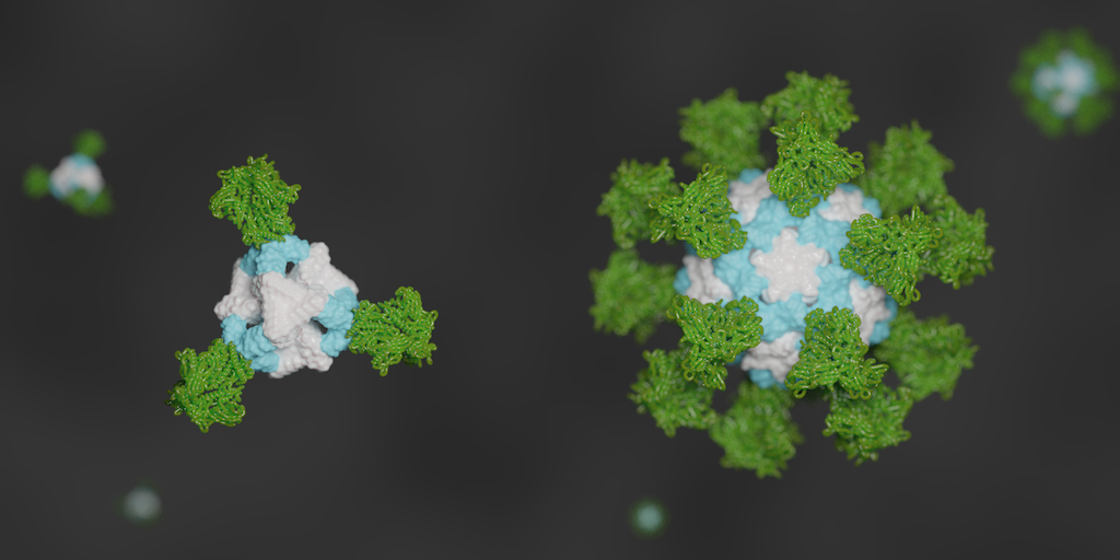

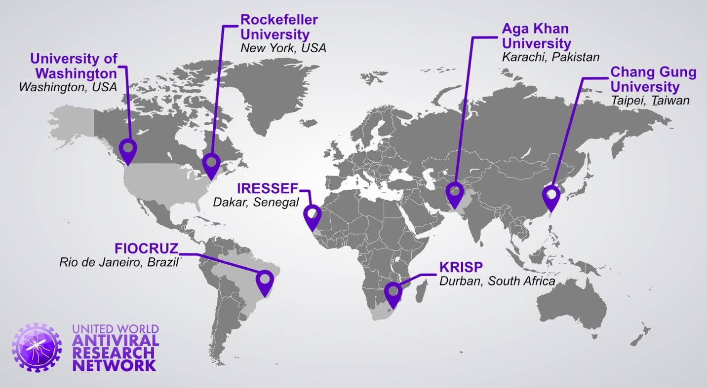
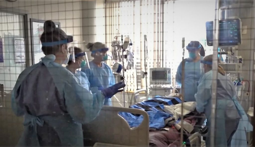
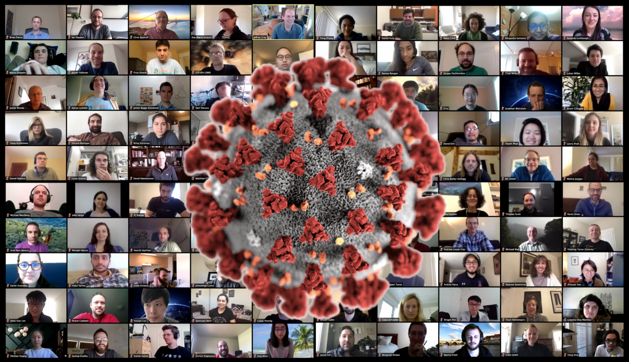
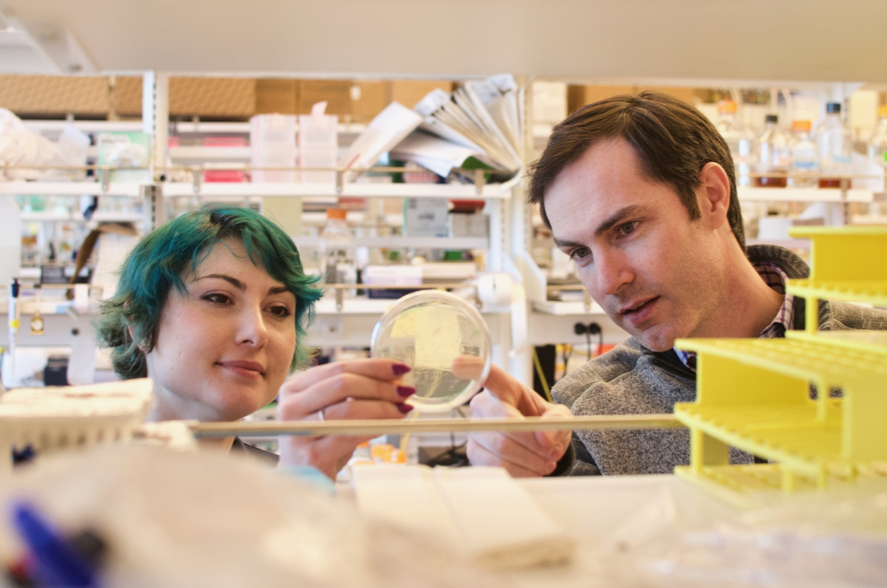
 The projections of 1 year to 18 months to have an FDA approved vaccine are probably accurate — if some of the vaccine candidates that [are] either in development or about to start clinical trials as of today do in fact provide protection (which we don’t know yet) and everything goes well.
 One long-term goal (which will not be ready for this pandemic) would be to create vaccines that could provide universal coverage against any family of viruses that has a high chance of causing a pandemic. For example, one could imagine having a single vaccine that protects against all possible coronaviruses, another vaccine that protects all possible flu viruses, etc. Based on what we know about how the immune system responds to flu, a universal flu vaccine does look possible as there are pieces of flu that are conserved between all flu viruses that infect humans. Coronaviruses are less studied in this regard. Hopefully greater study of them in the coming years will show that such a vaccine could be possible.
  There are a variety of proteins present in coronaviruses, but the spike protein is the major vaccine and antibody target because it is present on the outside of virus and is the major protein that our immune systems target during natural infection. The spike protein is also the workhorse of viral entry — the spike protein’s role is to bind to host cells and fuse viral and host membranes to allow for infection. The spike is therefore the first target the immune system sees and acts against. This also means that if we can target antibodies to it, we could prevent viral entry into host cells completely and block infection (which is the ultimate goal!). The most promising target on the spike protein is called the receptor binding domain, the goal being to block the interaction with host (our) receptor. If this interaction is blocked, then the virus cannot enter cells and no infection can occur.
 This is a really complex question, and we’ll only be able to provide a partial answer. If you are curious about how vaccines and drugs are developed, here are a few other resources:
  A few different vaccine candidates from other groups have entered the earliest stage of clinical testing, and many others are racing to get there. A handful of patients have been injected in Seattle as part of one mRNA vaccine trial (Phase 1).